The Ulutas Medical Journal · ISSN 2149-0430 (مطبوع), 2149-388X (إلكتروني) · https://www.ulutasmedicaljournal.com/?mno=8217
مقالة أصلية · وصول مفتوح
Retrospective Evaluation of the Role of CA19-9, CEA and Hematological Parameters in the Prediction of Helicobacter Pylori, Gastritis Etiology and Endoscopic Lesions
ترجمة آلية للعنوان والملخص والنص الكامل. الأصل الإنجليزي
- Department of Internal Medicine, Health Ministry, Bolu İzzet Baysal State Hospital, Bolu, Turkey ROR
- Department of Internal Medicine, Bolu Abant İzzet Baysal University, Faculty of Medicine, Bolu, Turkey ROR
الملخص
Introduction: Helicobacter pylori (HP) is an infectious disease that generates chronic gastric and duodenal mucosal tissue inflammation. أما نسبة النيوتروبول - ليمفوسيتي (NLR) ونسبة اللمفوسيت - الهضبة (PLR) فهي مؤشرات سهلة للالتهاب مصممة من الأشعة. واستهدفنا معرفة ما إذا كانت هناك صلة بين فيروس نقص المناعة البشرية والآفات الفموية الغازية، والأشعة الليبري، والبارامترات الوراثية، وعلامات الورم التي يمكن استخدامها لفحص السرطانات الشائعة في السكان، مثل مضادات الكربوهيدرات 19-9 (CA19-9) ومضادات الكاربونية. المواد والأساليب: ضمنا 100 من الذكور و 106 من الإناث الذين تقدموا بشكاوى مشبوهة إلى عيادات المرضى الخارجيين في المستشفى وخضعوا للنسخة العليا. جميع البارامترات من نظام تسجيل المستشفى تم تحليلها بأثر رجعي النتائج: لم يكن هناك فرق بين الفئات (-) السلبية (-) والسلبية (بنسبة 64 (31 في المائة) من الفئة HP الإيجابية (+) و 142 (69 في المائة). CA 19-9, NLR, PLR, and neutrophil count were higher in patients with gastric erosion than in patients with normal mucosa in the HP(+). PLR was higher in patients with gastric erosion, and leucocyte and monocyte counts were higher in patients with gastric ulcers than in patients with normal gastric mucosa in the HP(-) group. NLR and PLR were higher in the gastric erosion group than in the normal mucosa group, independently from HP. وإحصاءات النواة أعلى في المرضى الذين يعانون من السرطانات الغازية منها في المصابين بتآكل الغازات والسرطانات الغازية العادية. الاستنتاج: ولم يتمكن أي من هذه العلامات من التنبؤ بوجود الشرطة. وكانت الاستجابة المناعية، التي نجمت عن الالتهاب الناتج عن ضغط الدم، غير متجانسة؛ وقد تكون العوامل الوراثية وسلوك التغذية المختلفة أسبابا للحصول على بيانات مختلفة من هذه الدراسة وغيرها من الدراسات المماثلة بشأن هذه المسألة.
الكلمات المفتاحية
النص الكامل
ترجمة آلية للنص الكامل؛ النسخة المعتمدة هي الأصل الإنجليزي. الأصل الإنجليزي
مقدمة
ويؤثر مرض الداء، الذي يوصف بأنه ألم وقلق في البطن الأعلى، على نحو ربع السكان كل سنة. Although organic causes such as gastroduodenal ulcer, esophagitis, pancreaticobiliary disease, gastritis, duodenitis, gastric precancerous lesion, and/or gastric cancer can cause it, no underlying organic cause is frequently discovered.
والفحص الإندوسي هو الإجراء التشخيصي الأكثر شيوعاً لتحديد الأسباب العضوية. إن التهاب الغازي هو إلهاب المكوزا الغازي، الذي هو السبب الرئيسي للتشرد. According to the Sydney system of classification, it is divided into acute, chronic, and special forms, with special forms also accepted as chronic gastritis. HP is a significant human pathogen that causes peptic ulcer disease, gastritis, gastric adenocarcinomas, and gastric lymphoma, as well as dyspepcy (1, 29, 30)وعلى الرغم من أن البكتريوم لا يسبب سوى تهاب طفيف في كثير من الناس، فإن الالتهاب الغازي الذي يسببه ضغط الدم في الأجل الطويل يؤدي عادة إلى الالتهاب الفلكي بالغاز، الذي يُعتقد أنه المرحلة الأولى من مرض السرطان الغازي. (2, 31, 32)واستناداً إلى النتائج الوبائية، صنفت منظمة الصحة العالمية برنامج HP كعامل سرطاني من النوع الأول في عام 1994، وتم تأكيد هذا التصنيف في عام 2012. (3)- يمكن استخدام علامات الورم، مثل CA 19-9 و CEA، التي هي علامات بيولوجية يمكن استخدامها لفحص الخيول الشائعة في السكان، لفحص السرطانات الشائعة في الجمهور. CA 19-9 is increased in many types of gastrointestinal malignancies, including colorectal, hepatocellular, and esophageal cancers, and it is used to follow-up pancreatic cancer patients (4)- تستخدم الوكالة الدولية للطاقة الذرية، ولا سيما المرتبطة بالكولون أو الرئة أو الغدة الدرقية الدوائية أو الثدي أو الغازية أو الكبريتية، في متابعة سرطانات الألوان.(5)- تم تقييم البارامترات المغناطيسية كوسيلة لتقييم الالتهاب المنهجي في العديد من الأمراض. Since leukocytes induce an increase in neutrophil count and a decrease in lymphocyte count as a physiological response to stress, NLR and PLR can be evaluated as sensitive markers of systemic inflammation(6, 7) وكان الغرض من هذه الدراسة هو معرفة ما إذا كانت هناك أي صلة بين عدوى فيروس نقص المناعة البشرية والآفات الوراثية الغازية، و CA19-9، و CEA، و NLR، و PLR، والبارامترات الوراثية.
المواد والأساليب
المواضيع
وشملت الدراسة 100 من الذكور و 106 من الإناث، و 206 من المرضى الذين تجاوزوا سن 18 عاما الذين تقدموا بطلبات إلى عياداتنا الخارجية في الفترة ما بين حزيران/يونيه وتموز/يوليه 2018 بشكاوى مشبوهة، وخضعوا لنظام إندوسكوبيني أعلى من البنزين بعد الإعداد المناسب، وكان لديهم في نفس الوقت CA19-9، و CEA، وبارامترات مستمدة من الأشعة. وأُجريت الدراسة كدراسة استعراضية شاملة لعدة قطاعات.
البيان الأخلاقي
وقد وافقت مجالس الأخلاقيات المحلية على هذه الدراسة (2019/22) وتمتثل لأحكام إعلان هلسنكي الذي يعلن المبادئ الأخلاقية لتوجيه البحوث الطبية. وتم الحصول على موافقة مستنيرة من جميع المرضى قبل إجراء الإندوسكوبي.
الطرائق
وأُجريت عمليات النسخ باستخدام جهاز Olympus EVIS EXERA، GIF Q150 (Serial no: 2205164) . وسجل أخصائي علم الوراثة الوبائية التشخيصات الإندوسكوبية وفحص عينات الخزعة الإندوسكوبيكية عن طريق أخصائي علم الأمراض. وقد قُيِّمت مستويات نظام سيروم CEA ورقم CA 19-9 باستخدام أداة " أبوت التجارية " (Abottt Architect i2000SR) (مختبرات آبوت، بارك، IL، الولايات المتحدة الأمريكية). وتعتبر مستويات الوكالة من 0-3 نانوغرام/مليلتر و 19-9 بين 0-37 نانوغرام/مليلتر طبيعية. The hemogram was measured using the Abbott Cell-Dyn 3700 complete blood count tool (Abbott Laboratories, Abbot Park, IL, USA), which examined cells in three dimensions using laser light scattering and fluorescent signals from two angles. وقد حُسبت نتائج الأشعة فوق البنفسجية والأشعة فوق البنفسجية. وتم الحصول على جميع البيانات المسجلة بأثر رجعي باستخدام نظام تسجيل المستشفيات.
المرضى الذين تقل أعمارهم عن 18 سنة، والحوامل أو المرضعات، والتهاب الكبد الحاد أو المزمن، والتهاب الكبد، والتهاب الكبد الحاد، والتهاب الكبد الحاد، وداء الجوند المعوي، ومرض الأمعاء الهادف، والنزيف الفعّال من نوع GI، وتاريخ التهاب السكري غير الخاضع للمراقبة، وارتفاع ضغط الدم، والعدوى الرئوية، والعدوى الطويلة الأجل2 وقد استبعدت من هذه الدراسة مصدِّق أو استخدام مضادات حيوية في الأسبوعين الماضيين، وعرقلة و/أو ارتجاج نظم المعلومات الجغرافية، والمدخنين النشطين، والمتلقين للعلاج باستئصال شأفة الكبد.
التقييم الإحصائي
For statistical analysis, IBM SPSS 20 (Statistics Program for Social Scientists) was used. To evaluate whether the distribution is normal, Kolmogorov-Smirnov test, couplewise comparison of continuous variables, Student t-test for those with normal distribution, Mann-Whitney-U test for those without normal distribution, multiple comparisons of continuous variables, One-Way ANOVA for those with normal distribution, Kruskal-T for those without normal distribution. واستُخدمت اختبارات (واليس) واختبارات (تشيسكوير) واختبارات (فيبر) بدقة لمقارنة البيانات القاطعة. وقُبل مستوى أهمية الفقرة 10-5.
النتائج
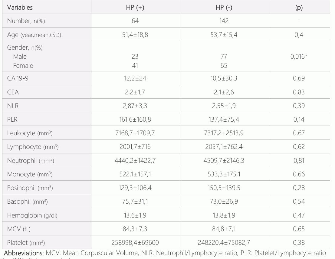
وكان متوسط عمر 206 مريضاً مدرجاً في الدراسة هو 5316.5 مريضاً، و64 مريضاً (31 في المائة) من المصابين بفيروس نقص المناعة البشرية (+). وكان أصغر سن هو 18 عاما، بينما كان أقدم العمر 83 عاما. وكان متوسط عمر المرضى (+) 51.4 18.8 سنة و 41 (64 في المائة) من النساء و 23 (36 في المائة) من الرجال. وكان متوسط العمر لـ (-) 53.715.4 سنة، و65 (45.7 في المائة) للنساء، و77 (54.2 في المائة) رجال [الجدول 1]. وفي كلتا الفئتين، لم يكن هناك فرق في العمر. (ع=0.40). وقد تبين أن عدد الأفراد من الفئة " HP " (+) أعلى من عدد الذكور من الناحية الإحصائية (p=0.016). In terms of CA19-9, CEA, NLR, PLR, and other hemogram parameters, there was no significant difference between individuals with HP (+) and (-).
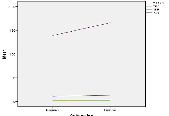
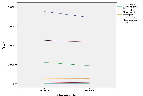
The data were analyzed for the antrum and corpus based on the presence of HP. According to the presence of HP in the gastric antrum and also corpus region, the distribution of serum CA 19-9, CEA, NLR, PLR levels was divided into two groups as HP(+) and HP(-). In terms of serum CA 199, CEA, NLR, and PLR levels, there was no significant difference between these groups [Figure-1]. As a result of endoscopic biopsy and histopathological evaluation of gastric corpus region, your were divided into two groups as HP(+)/(-) and hemogram parameters such as leukocyte, lymphocyte, monocyte, neutrophil, basophil, eosinophil, hemoglobin and MCV. The lymphocyte count in the corpus was found to be significantly higher in those with HP(-) compared to HP(+) (p=0.018, 2228 744.7 vs. 1868650.9) [Figure-2].
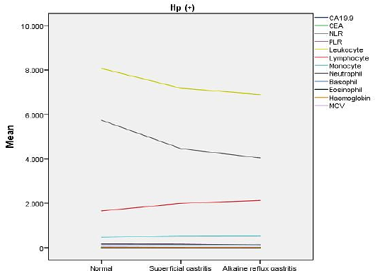
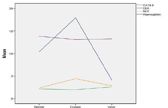
When the patients were evaluated according to their gastritis status as a result of endoscopy, gastric mucosa was found normal in 8 patients, superficial gastritis in 162 patients, and alkaline reflux gastritis in 36 patients. When HP (+) individuals with normal gastric mucosa, those with superficial gastritis and those with alkaline reflux gastritis were analyzed according to serum CEA, CA 199, NLR, PLR levels and the other hemogram parameters, no difference was found between individuals with normal gastric mucosa, those with superficial gastritis and alkaline refluxtri. As a result of their endoscopic evaluation, the patients were divided into three groups those with normal gastric mucosa, those with gastric erosion and those with gastric ulcer, and also were evaluated according to CA 199, CEA, NLR levels and the other hemoglobin parameters. The NLR was higher in the gastric erosion than in the normal gastric mucosa (p=0.0001). The levels of serum CA19-9, CEA, and hemoglobin did not differ significantly between the groups (p.0.05) [Figure-4].
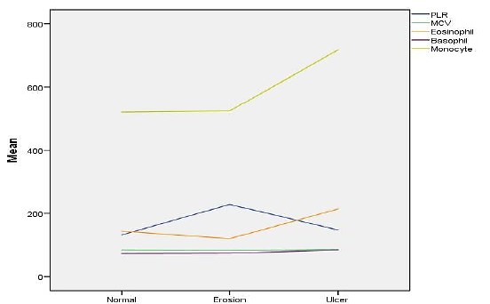
When the patients were divided into three groups as those with normal gastric mucosa, those with gastric erosion and those with gastric ulcer as a result of their upper GIS endoscopic evaluation, they were evaluated according to serum PLR, MCV, eosinophil, basophil and monocytes levels. وقد تبين أن مستوى PLR أعلى في مجموعة التعرية الغازية منه في المجموعة العادية للغاز (p=0.0001). ولم يكن هناك أي فرق كبير بين المجموعات من حيث مستويات الأشعة المقطعية والنسيج والبازوفيل والاحتكار. وقد تبين أن معدل احتكار السرطان أعلى بكثير في مجموعة السرطانات الغازية مقارنة بالتضخم الغازي الطبيعي والتحات الغازي (p=0.002, p=0.008, respectively) [Figure-5].
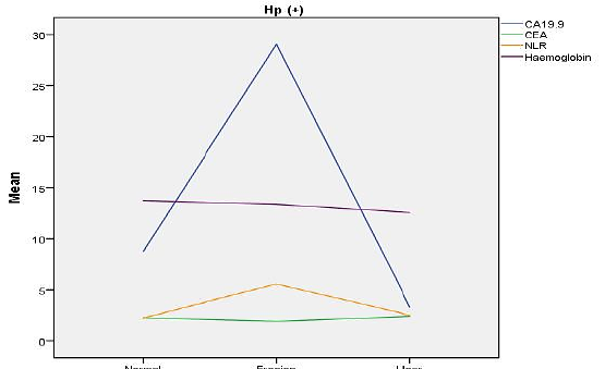
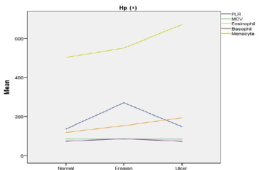
When the individuals with HP (+) were divided into 3 groups as those with normal gastric mucosa, those with gastric erosion and those with gastric ulcer according to the results of upper GI endoscopy, the CA19-9 and NLR levels were higher in the group with gastric erosion compared to the normal gastric mucosa (p= 0.021, p=0.004, respectively). ولم يكن هناك فرق بين المجموعات من حيث المصل CEA ومستويات الهيموغلوبين [الشكل 6]. When individuals with HP (+) were divided into 3 groups as those with normal gastric mucosa, those with gastric erosion and with gastric ulcer according to the results of upper GI endoscopy, the PLR was higher in the gastric erosion than in the normal gastric mucosa group (p=0.024). There was no difference between the groups in terms of MCV, eosinophil, basophil and monocyte levels [Figure-7].
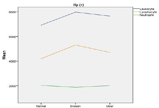
When individuals with HP(+) were divided into 3 groups as those with normal gastric mucosa, those with gastric erosion and those with gastric ulcer according to the results of upper GI endoscopy, the neutrophil count was found to be higher in the gastric erosion group than in the normal gastric mucosa group (p=0.038). In terms of serum leukocyte and lymphocyte levels, there was no difference between the groups [Figure-8].
المناقشة
واحدة من أكثر الشكاوى المعروفة في نظام المعلومات الجغرافية هي التشريح. وهي أكثر شيوعا بين الرجل والمرأة، وتزداد حالاتها مع بلوغها سنا. ولوحظ أن انتشار الإصابة بالمرض بين 25 و5 في المائة في مجتمعات مختلفة (8)The Gastritis, duodenal ulcer, gastric ulcer, GERD, NSAID use, and antibiotic use are among the most common causes of organic dyspepsia seen in the clinic (9)وتفيد التقارير بأن الإصابة بفيروس نقص المناعة البشرية أكثر شيوعاً في مرضى مرضى مرضى مرضى مرضى الداء بدلاً من وجود ضوابط صحية. (10)- إن الإصابة المزمنة بفيروس نقص المناعة البشرية شائعة في نبرة المعدة في البلدان المتقدمة النمو، وهي ترتبط عادة بالسرطانات الدموية. In developing countries, HP infection is more localized in the corpus. In this case, HP infection is the initiator of the cascade of chronic gastritis, gastric atrophy, intestinal metaplasia, dysplasia, and invasive carcinoma (11)- على الرغم من أن الدراسات تُقيِّم العلاقة بين مؤشري الورم وعلامات الورم والبارامترات المغناطيسية (6, 12, 13)ولا توجد بيانات كافية في الأدبيات المتعلقة بالدراسات الأخيرة التي تحقق في العلاقة بين التهاب البنزين السطحي، والتلوث بالغاز الألكيليني، وتآكل السرطان الغازي، وعلامات الورم والبارامترات الوبائية. وقد ألهمتنا القدرة على التنبؤ بالمرضيات الغازية التي تشخيصها بعض الإجراءات مثل الاستنساخ، مع علامات يمكن رؤيتها في المصل، إلى العمل في هذا الاتجاه. وتظهر الدراسات المختلفة في الأدبيات أن تواتر هذا البرنامج يختلف حسب السنوات والمناطق، وهو أقل نسبيا في المجتمعات الغربية وغرب تركيا. (14–18) - وقد أدت بعض العوامل، مثل عدم كفاية مستوى ضغط الدم في البيئة، والتوزيع الرقيقي للحمض الغازي، والتحقيق في وجود فيروس نقص المناعة البشرية باستخدام مرض التهاب الكبد باعتباره الطريقة الوحيدة، إلى أن يكون تواتر ضغط الدم في مجموعتنا المريضة أقل من النتائج التي خلصت إليها الدراسات السابقة.
وفي هذه الدراسة، وجدنا أنه لم يكن هناك فرق كبير بين مستويات CA 19-9 و CEA في مجموعات HP (+) و (-). In the study of Xu et al. (12)(أ) كان مستوى نظام المحاسبة البيئية - الاقتصادية الذي وجد في مَن لديهم (+) و(-) كبيراً (p0.001). وثمة سبب هام للاختلافات في نتائج هذه الدراسة هو أن النتائج قد تكون أكثر جدوى بسبب الاختلافات الاجتماعية - الثقافية والعادات التغذوية لسكان هذه الدراسة والأفراد الصينيين، وأن حجم العينة في الدراسة أكبر بكثير. ومع ذلك، فإن ما يجعل نتائج هذه الدراسة أكثر قيمة هو التشخيص المرضي للإصابة بفيروس نقص المناعة البشرية كأسلوب. Xu et al. used only serology for diagnostic purposes. The specificity of histopathology (95-98%) in the diagnosis of HP infection is higher than the specificity of serological tests (86-95%). In terms of CA 19-9 levels, no significant difference was found in terms of HP infection in both studies.
In inflammatory processes and infections, leukocyte, neutrophil, and the platelet count can increase. وينشط برنامج HP الآليات التي تعمل وتمنع الاستجابة المناعية. In HP, leukocytosis, neutrophilia, monocytosis and basophilia are expected as a systemic effect of mucosal inflammation. In the present study, leukocyte and neutrophil counts were found to be statistically insignantly higher in the HP(-) group (p=0.67, p=0.81)(19)ونظراً لأن عدد مركبات ثلاثي بوتيل القصدير يمثل أيضاً تفاعلاً حاداً في المرحلة، فمن المتوقع أن يزيد من عمليات التهاب مثل عدوى فيروس نقص المناعة البشرية. It causes an increase in MPV (mean platelet volume), PDW (platelet distribution width), PLR (platelet/lymphocyte ratio) and PLT numbers (20).
In various studies, when HP (+) and (-) patients are compared, it is seen that different results are obtained in terms of hematological parameters. والسبب في النتائج التي تتمخض عنها هذه الطريقة هو أن اليقظة البكتيرية واستجابة نظام الدفاع المضيف تظهر اختلافات كبيرة بين شخص وآخر، وهذا ينعكس في القيم المختبرية. ولم نتمكن من الحصول على بارامتر ذي مغزى يرشدنا في تشخيص وتشخيص الإصابة بفيروس نقص المناعة البشرية، ولا سيما من صيغة اللوكوست. وكانت النتيجة التي توصلنا إليها في الدراسة التي تفيد بأن معدلات الإصابة بفيروس نقص المناعة البشرية/الإيدز ومعدلات الإصابة بفيروس نقص المناعة البشرية أعلى في مرضى فيروس نقص المناعة البشرية (+) متمشية مع الأدبيات التي تفيد بأن هذه البارامترات هي علامات موثوق بها للتهاب. غير أن الافتقار إلى الأهمية الإحصائية لنتائجنا لا يسمح لنا بتقديم توصيات بشأن استخدام هذه البارامترات في تشخيص ومتابعة الإصابة بفيروس نقص المناعة البشرية (20–22, 27, 28).
The HP pathogen exhibits different behavioural modalities in the gastric antrum and corpus. In the gastric antrum, it is mostly associated with ulcers, whereas in the gastric corpus region it is more carcinogenic and has a stronger association with gastric cancer. لذلك، قيّمنا نتائج خزعة الغدد الصماء للمرضى بشكل منفصل من أجل النتروم والجثة. In the antrum, there was no difference between the groups with HP(+) and HP(-) and CEA, CA19-9, NLR, PLR and hemogram parameters. The lymphocyte count in the corpus was found to be higher in the HP (-) than in the HP (+) group. (p=0.018). وتشير الدراسات الواردة في المؤلفات إلى أن عدد اللمفوسيتي أعلى وأدنى على حد سواء في عدد الألف (+) (19, 22, 23).
There were no differences between CEA, CA 19-9 levels, NLR, PLR, and the other hemogram parameters when the groups with HP (+) and HP (-) were evaluated separately according to the presence of gastritis [Figure-3].
The NLR and PLR values were higher in gastric erosion than in normal gastric mucosa in the present study (respectively p=0.0001, p=0.0001) [Figure-4, 5]. While the NLR and PLR elevations in those with gastric erosion in the HP (+) group remained significant (p=0.04, p=0.024), in the HP(-), although the NLR elevation in those with gastric erosion lost its significance, PLR elevation was higher than the normal gastric mucosa maintained its significance (p=0.011). وكان للمرضى المصابين بالسرطانات الغازية عدد أعلى من عدد المصابين بالسرطانات الغازية الطبيعية والتحات الغازي (من الناحية النظرية، ص = 0.002، ص = 0.008) [الشكل 5]. Although gastric ulcer is not considered a cause of monocytosis, monocyte levels were higher in patients with gastric ulcer compared with patients with normal gastric mucosa and gastric erosion in the present study. In the HP (+), those with gastric ulcer lost their monocyte uptrend compared to gastric erosion and normal gastric mucosa, whereas in the HP(-); those with gastric ulcers maintained significant monocyte elevation compared to gastric erosion and normal mucosa (p=0.014, p=0.011). وقد يكون ارتفاع مستوى احتكار الخادم البارامترات المفيدة في التنبؤ بالقرح الغازي. ولم تكن هناك دراسة أو استنتاج بشأن هذا الموضوع حدد في المؤلفات. وقد برزت ضرورة إجراء مزيد من البحوث بشأن هذا الموضوع.
وكانت مستويات الإصابة بفيروس نقص المناعة البشرية من 19 إلى 9 أعلى في مرضى فيروس نقص المناعة البشرية (+) الذين يعانون من تآكل غازي بالمقارنة مع المصابين بالسرطانات الغازية العادية [الشكل 6] (p=0.021). ولم يكن هناك فرق بين مستويات الوكالة. In the HP (+) group, CA 19-9 elevation may be an useful parameter in predicting erosion. ومع ذلك، فإن العدد القليل من المرضى في الدراسات التي تقترح هذه العلاقة، (24, 25) وتشير هذه الدراسة إلى ضرورة إجراء دراسات أكبر بشأن هذا الموضوع.
وقد تبين أن عدد نوتروفيل ومعدلات الإصابة بفيروس نقص المناعة البشرية/الإيدز أعلى كثيراً في مرضي HP(+) مع التحات الغازية (p=0.038, p=0.004, p=0.024, respectively) [Figure-6 and Figure-8]. وقد تبين أن معدل الإصابة بالعدوى النووية ومعدل الإصابة بالفيروس أعلى بكثير في المرضى الذين يعانون من تآكل الغاز في مجموعة HP (+) مقارنة بالمرضى الذين يعانون من تسمم غازي عادي في هذه الدراسة، وقد جرى تقييم كل منهما بصورة مستقلة عن وجود HP. According to these results, erosion-related inflammation that developed independently of the presence of HP may have been triggered, and accordingly, NLR and PLR levels, which were accepted as inflammation markers, might be significantly higher than those with normal gastric mucosa (20, 21, 26).
وبالنظر إلى موثوقية هذه الدراسة، يمكن ملاحظة أن المرضى الذين يعانون من ظروف متجانسة قد تؤثر على نتائج الدراسة مستبعدون من الدراسة، وأن مجموعة المرضى التي درست بهذه الطريقة أكثر تجانساً، وأن المعلومات تستند إلى سجلات موثوقة، وأن وجود فيروس نقص المناعة البشرية يكشف عن حدوث داء الغدد الصماء. وتتسم الدراسة ببعض القيود؛ وبما أن جميع الأفراد المدرجين في هذه الدراسة كانوا مرضى قدموا طلباً إلى كلية الطب في عيادة المرضى الخارجيين المشمولين بالشكاوى المشبوهة، فقد لم يكن كافياً لتمثيل عامة السكان. فحصنا التعاطفي كشف وجود الشرطة وحساسية هذا البرنامج بالنسبة لاستقطاب ضغط الدم تبلغ نحو 80 في المائة، ويعود ذلك إلى حد كبير إلى ضلوع ضغط الدم في المعدة. ولذلك، ربما نكون قد نظرنا في بعض HP(+) على أنه (-). ويمكن تخفيض هذا المعدل بإضافة اختبار ثانٍ مثل التشخيص والفحص الرئوي للتشخيص. The study may have been insufficient to represent larger groups since a total of 206 patients were evaluated because patients were selected according to certain exclusion criteria. وبما أن هذه الدراسة هي دراسة استطلاعية للمراقبة، فإنها لا تستطيع أن تقدم معلومات كافية من حيث الكشف عن أسباب تقييم النتائج للعلاقة بين الإصابة بفيروس نقص المناعة البشرية ونتائج الغدد الصماء وقيم ورم المصل، ومستويات الأشعة الليبري، والبارامترات الرئوية الأخرى. وعلى الرغم من أن الذين لديهم تاريخ من السرطان النشط أو السابق وأولئك الذين تم تشخيصهم بالسرطان نتيجة الاستنساخ لم يدرجوا في الدراسة، لا يمكننا تجاهل إمكانية وجود مرضى مصابين بالسرطان غير المشخص في عينتنا، وأن القيم المختبرية لهؤلاء الأفراد قد أثرت على نتائج هذه الدراسة.
خاتمة
وفي حين أن مصدر معظم حالات التشنج غير معروف، فإننا نعرف أن بعض المرضى لديهم شكاوى مشبوهة يمكن تشخيصها عن طريق فحص الغدد الصماء. وفي هذه الدراسة، نسهم في الأدبيات عن طريق التحقيق في إمكانية التنبؤ بالنتائج التي يتم التوصل إليها عن طريق التقييم الإندوسي بالمصل CA 19-9، ومستويات CEA، وبارامترات الأشعة، ونعتقد أنه سيكون مصدر إلهام لإجراء مزيد من الدراسات بشأن هذا الموضوع في تصميم أوسع نطاقا ومتوقعا.
الإقرارات
- مساهمات المؤلفين (CRediT)
- ساهم جميع المؤلفين بالتساوي في جميع أدوار CRediT الأربعة عشر (Conceptualization, Data curation, Formal analysis, Funding acquisition, Investigation, Methodology, Project administration, Resources, Software, Supervision, Validation, Visualization, Writing – original draft, Writing – review & editing).
- التمويل
-
Funding information was not reported in the published version of this article.
- تضارب المصالح
- All authors declare no conflict of interest.
- الموافقة الأخلاقية
- The present study was approved by the local ethics boards (2019/22) and complies with the provisions of the Declaration of Helsinki, which declares ethical principles to guide medical research. Informed consent was obtained from all patients before the endoscopic procedure.
- إتاحة البيانات
- A data availability statement was not reported in the published version of this article.
المراجع (32)
16 من أصل 32 مرجعًا مرتبطة بمعرّف DOI.
- Forman D. The prevalence of Helicobacter Pylori infection in gastric cancer. Aliment Pharmacol Ther. 1995;2:71-6 PubMed Google Scholar
- Nedrud JG, Slanchard SS, Czinn SJ. Helicobacter pylori inflammation and immunity. Helicobacter 2002;1: 24-9 DOI Crossref PubMed Google Scholar
- World Health Organization/International Agency for Research on Cancer: IARC Monographs on the Evaluation of Carcinogenic Risks to Humans. Vol 61: Schistosomes, Liver Flukes and Helicobacter pylori. Lyon, International Agency for Research on Cancer, 1994 PubMed Google Scholar
- Perkins G, Slater, E, Sanders, G, Prichard J. "Serum tumor markers". American Family Physician 2003;68(6):1075–1082. PubMed Google Scholar
- Goldstein MJ, Mitchell EP. "Carcinoembryonic antigen in the staging and follow-up of patients with colorectal cancer". Cancer Investigation 2005;23(4):338–51 DOI Crossref PubMed Google Scholar
- Farah R, Hamza H, Khamisy-Farah R. A link between platelet to lymphocyte ratio and Helicobacter pylori infection. J Clin Lab Anal. 2018;32: 1 DOI Crossref PubMed Google Scholar
- Lubrano V, Zym L, Balzan S. Neutrophil to lymphocyte ratio and c reactive protein are independent predictors of severe coronary artery disease: comparison with hydroperoxides and consolidated inflammatory markers. Theor Biol Forum. 2018; 111(1-2):49-56 PubMed Google Scholar
- Knill-Jones RP. Geographical differences in the prevalence of dyspepsia. Scand J Gastroenterology 1991;26:17-24 DOI Crossref PubMed Google Scholar
- Özden A, Şahin B, Yılmaz U et al. Dyspepsia, Helicobacter pylori. Gastroenterology, TGV publication. Ankara 2002:85-72 77 Hematological Parameters Taşçı et al. PubMed Google Scholar
- Armstrong D. Helicobacter pylori infection and dyspepsia. Scand J Gastroenterol Suppl. 1996;215:38 -47 DOI Crossref PubMed Google Scholar
- Smith VC, Genta RM. Role of Helicobacter pylori Gastritis in Gastric Atrophy, Intestinal Metaplasia, and Gastric Neoplasia. Microscopy Research and Technique 2000;48:313-20 DOI Crossref PubMed Google Scholar
- Xu MY, Cao B, Chen Y,et al. Association between Helicobacter pylori infection and tumor markers: an observational retrospective study. BMJ Open. 2018;8(8). DOI Crossref PubMed Google Scholar
- Yıldız R, Cindoruk M, Büyükberber S, et al. CA 19-9, CEA and CA 72-4 levels in erosive gastritis, Gazi Medical Journal, 2009;20(2):55-56 PubMed Google Scholar
- The Eurogast Study Group. Epidemiology of and a risk factor for Helicobacter pylori infection among 3194 asymptomatic subjects in populations. GUT 1993;34(12):1672-6 DOI Crossref PubMed Google Scholar
- Özardalı Hİ, Bitiren M, Nazlıgül Y, Yılmaz N. The frequency of Helicobacter pylori in non-erosive gastritis in Şanlıurfa region. Journal of General Medicine 1998;8(1):149-52 PubMed Google Scholar
- Konakcı N, Gülten M, İbanoğlu MS, et al. The frequency of Helicobacter pylori in patients with chronic active gastritis. Journal of Uludağ University. 2010;36(1):7 -10 PubMed Google Scholar
- Joos A, Nemzeth A, Zsolnay G, et al. Gastric biopsies and Helicobacter pylori. ORV Hetil 1995;136(12):1975 -82 PubMed Google Scholar
- Turan G, Altun E. "The frequency of Helicobacter pylori in endoscopic biopsies with gastritis findings in Balıkesir region". Balıkesir Medical Journal. 2017:17-21 PubMed Google Scholar
- Karttunen TJ, Niemelä S, Kerola T. Blood leukocyte differential in Helicobacter pylori infection. Digest Dis Sci 1996;41,1332-36 PubMed Google Scholar
- Kaplan M, Ateş İ, Yüksel M, et al. The role of the PLR–NLR combination in the prediction of the presence of Helicobacter pylori and its associated complications. Saudi J Gastroenterol 2018;24:294-300 DOI Crossref PubMed Google Scholar
- Farah R, Farah RK. Association of Neutrophil to Lymphocyte Ratio With Presence and Severity of Gastritis Due to Helicobacter pylori Infection. Journal of Clinical Laboratory Analysis, 2014;28:219–23 DOI Crossref PubMed Google Scholar
- Zeren S, Bayhan Z, Koçak FE, et al. Diagnostic value of platelet/lymphocyte ratio and neutrophil/lymphocyte ratio in investigations for helicobacter pylori gastritis. Int J Clin Exp Med 2016;9(2):5102-6 PubMed Google Scholar
- Güçlü M, Agan AF. Association of Severity of Helicobacter pylori Infection with Peripheral Blood Neutrophil to Lymphocyte Ratio and Mean Platelet Volume. Euroasian J Hepatogastroenterol. 2017;7(1):11–16 DOI Crossref PubMed Google Scholar
- Paul A, Bunn JR, Cohen MI, et al. Simultaneous Gastric and Plasma Immunoreactive Plasma Carcinoembryonic Antigen in 108 Patients Undergoing Gastroscopy Gastroenterology, 1979;76:734-741 DOI Crossref PubMed Google Scholar
- Ventrucci M, Pozzato P, Cipolla A, Uomo G. Persistent Elevation of Serum CA 19-9 With No Evidence of Malignant Disease. Dig Liver Dis. 2009;41(5):357-63 DOI Crossref PubMed Google Scholar
- Jafarzadeh A, Akbarpoor V, Nemati M, Rezayati MT. Total Leukocyte Counts and Neutrophil-Lymphocyte Count Ratios Among Helicobacter Pylori-Infected Patients With Peptic Ulcers: Independent of Bacterial CagA Status. Southeast Asian J Trop Med Public Health. 2013;44(1):82-8 PubMed Google Scholar
- Gasparyan AY, Ayvazyan L, Mukonova U, et al. The Platelet-to-Lymphocyte Ratio as an Inflammatory Marker in Rheumatic Diseases. Ann Lab Med. 2019; 39(4):345-57 DOI Crossref PubMed Google Scholar
- Pan L, Du J, Li T, Liao H. Platelet-to-lymphocyte Ratio and Neutrophil-To-Lymphocyte Ratio Associated With Disease Activity in Patients With Takayasu's Arteritis: Case-Control Study. BMJ, 2017;7(4). DOI Crossref PubMed Google Scholar
- The EUROGAST Study Group. Gut 1993;34:1672-6 PubMed Google Scholar
- Malaty HM, Engstrad L, Pedersen NL, Graham DY. Helicobacter pylori infection. Genetic and environmental influences. A study of twins. Ann Intern Med 1994;120:982-6 DOI Crossref PubMed Google Scholar
- Helicobacter pylori. Biologic Agents: A Review of Human Carcinogens, vol 100B. Lyon, International Agency for Research on Cancer. 2012;385-435 PubMed Google Scholar
- Di Napoli A, Petrino R, Boero M, et alınarak. Quantitive assessment of histologic changes in chronic gastritis after eradication of Helicobacter pylori. J Clin Pathol 1992;45:796-8 PubMed Google Scholar
اقرأ النص الكامل
افتح ملف PDF هنا
جارٍ تحميل ملف PDF…
إذا لم يظهر ملف PDF هنا، استخدم زر التنزيل أعلاه.
كيف تقتبس هذه المقالة
Tasci B, Gurler M. Retrospective Evaluation of the Role of CA19-9, CEA and Hematological Parameters in the Prediction of Helicobacter Pylori, Gastritis Etiology and Endoscopic Lesions. Ulutas Med J. 2022;8(1):64-72. doi:10.5455/umj.20220118095245
Tasci, B., & Gurler, M. (2022). Retrospective Evaluation of the Role of CA19-9, CEA and Hematological Parameters in the Prediction of Helicobacter Pylori, Gastritis Etiology and Endoscopic Lesions. The Ulutas Medical Journal, 8(1), 64–72. https://doi.org/10.5455/umj.20220118095245
Tasci, B. and Gurler, M. (2022) 'Retrospective Evaluation of the Role of CA19-9, CEA and Hematological Parameters in the Prediction of Helicobacter Pylori, Gastritis Etiology and Endoscopic Lesions', The Ulutas Medical Journal, 8(1), pp. 64–72. doi: 10.5455/umj.20220118095245.