The Ulutas Medical Journal · ISSN 2149-0430 (مطبوع), 2149-388X (إلكتروني) · https://www.ulutasmedicaljournal.com/?mno=282591
بحث أصلي · وصول مفتوح
A Blocking Barrier During Reduction of the Elbow Dislocation in Children: Incarcerated Medial Epicondyle Fracture
ترجمة آلية للعنوان والملخص والنص الكامل. الأصل الإنجليزي
- Orthopaedics and Traumatology, Ankara Numune Training and Research Hospital, Ankara, Turkey ROR
الملخص
معلومات أساسية: في الأطفال، قد يغيب عن الأشعة التقليدية كسر ملحمي وسطي في الركاز المرتبط بتفكك القوس. وهناك معايير إشعاعية أخرى موثوقة لا بد منها لتشخيص هذه الكسور. The purpose of the study was to investigate the clinical results of the operative treatment of incarcerated medial epicondyle fractures after elbow dislocation in children and define new radiological criteria in the diagnosis. المواد والأساليب: رُئي أن خطا مزدوجا يشبه موجة البحر يتكون من خطين متوازيين يتكونان من موجات ضوئية من المصابين بأمراض الأطفال غير المكتظة. وعُرِّف تعطيل اللافتة المشابهة للموجات البحرية بأنه معيار تشخيصي جديد في الكسور الوسيطة المسجونة بعد تشتيت القوس في الأطفال. سبعة أطفال مع كسر ملحمي وسطي مسجون النتائج: بلغ متوسط العمر 12.142.1 سنة (من 9 إلى 15 سنة) وكان متوسط فترة المتابعة 29.113.6 شهر (من 12 إلى 48 شهرا). The mean Mayo elbow score was 92.84.8 (range: 85-100) at the final follow-up. وتم تحديد المعيار التشخيصي الجديد للإشعاع في جميع المرضى المكسورين في الوسط. الاستنتاجات: The results of this study suggest that open reduction internal fixation of incarcerated medial epicondyle fractures after elbow dislocation resulted in satisfactory motion and function. والمعيار الإشعاعي التشخيصي الجديد الذي حددته هذه الدراسة مفيد، ولا ينطوي على أي تكلفة إضافية وقد يحل مشاكل التشخيص التي يعاني منها الكسور الوبائية المتسربة في الركاز بعد تشتيت القوس في السكان الأطفال.
الكلمات المفتاحية
النص الكامل
ترجمة آلية للنص الكامل؛ النسخة المعتمدة هي الأصل الإنجليزي. الأصل الإنجليزي
مقدمة
كسور ملحمية وسيطة تغطي ما يصل إلى 20 في المائة من كسور الولادة (1)- ما زالت إدارة الكسور المنعزلة والمشردة للوسط الرطبة تثير الجدل. وقد دُعي إلى التعبئة والنُهج الجراحية على حد سواء. تم الإبلاغ عن نتائج جيدة مع العلاج غير التشغيلي، على الرغم من ارتفاع معدل عدم الشمل (2–5)- في حالة المرضى الذين يعانون من كسور مشردة، توفر النُهج الجراحية الاستقرار والنطاق الوظيفي للحركة.
أحياناً، الشظية المكسورة قد تحتل الشظية الخلفية The presence of dysesthesias in the territory of the ulnar stress requires urgent open reduction of the incarcerated fragment. In children, an incarcerated epicondyle fracture of the distal humerus associated with elbow dislocation may be missed on diagnosed on conventional radiographs. وهناك معايير إشعاعية أخرى موثوقة لا بد منها لتشخيص هذه الكسور.
The purpose of the study was to investigate the clinical results of the operative treatment of incarcerated medial epicondyle fractures after elbow dislocation in children and to define a new radiological criterion in the diagnosis.
المواد والأساليب
وقد أُدير ما مجموعه 30 مريضاً يعانون من كسور ملحمي في وسائط الإعلام النازحين بالتخفيض والتثبيت في مؤسستنا في الفترة 2007-2015. وأُجري استعراض للحالة الرجعية لـ 7 أطفال متتاليين يعانون من كسر ملحمي وسطي مسجون بعد تشتيت القوس. وقد وافق مجلس الاستعراض المؤسسي على الدراسة. The inclusion criteria of this study were that patients were younger than 16 years, and had an incarcerated medial epicondyle fracture with at least a 12-month follow-up period. وقد استُبعدت من الدراسة المرضى الذين يعانون من أي تشوهات ضارة في النسل الأعلى أو من الجراحة السابقة للمرفق.
وتم تقييم جميع المرضى بالأشعة الفوقية والخلفية التقليدية. ولتيسير تشخيص الكسور الوسيطة المسجونة، حددنا معايير تشخيصية جديدة. A parallel double the line was seen as a ‘sea wave-like sign on the anterior-posterior elbow radiographs of non-fractured or reduced pediatric patients (Figure- 1). This parallelism was seen to be disrupted in incarcerated medial epicondyle fractures. The incarceended fragmented in the joint space in the medial compartment of the elbow between the trochlea of the humerus and coracoid of the ulna leads to disruption of this parallel double line. وقد حددنا هذا التعطل للعلامة المشابهة للموجات البحرية كمعيار إشعاعي تشخيصي جديد للكسرات الوسيطة المسجونة بعد تشتيت القوس في الأطفال (الشكل 2).
جميع المرضى خضعت لعملية جراحية باستخدام تقنية مماثلة المرضى كانوا يرتدون حساء تمّ استخدام مُعدّل مغناطيسي. المرضى كانوا مستعدين ومعالجين بطريقة عادية The affected arm was externally rotated with the elbow in a 90° - 100° flexed position. تم شق جلدي بحوالي 5 سنتيمتر على شرفة الوسط وطبق تفكك عميق ووُقعت الأعصاب الرئوي. وتم العثور أيضاً على الإلحاح الوسيط. The fragment was reduced with the arm in flexion and fixed with two Kirschner wires. واستُخدمت رسومات إذاعية لتأكيد الخفض ومواقع كيرشنر اللاسلكية. تم تقييم استقرار القوس تم تطبيق إغلاق الجلد
وبعد التشغيل، استُخدِمَت مجموعة طويلة من الأسلحة لمدة ثلاثة أسابيع. The range of motion of the elbow was started after cast removal. أزيلت أسلاك كيرشنر بعد 4-6 أسابيع ولا تطبق العلاج الفيزيائي إلا إذا كان هناك انخفاض ملحوظ في حركة القوس بعد 6 أسابيع من ترحيله. وكان جميع المرضى متابعين منتظمين في 3 و 6 و 12 و 24 و 36 شهرا بعد التشغيل. وأُجريت دراسة قياسية للأشعة الأمامية والأفقية للمرفق في فحص المتابعة، وقُيِّمت مضاعفات مثل مشاكل الجروح.
النتائج
وكان عدد المرضى 6 ذكور و 1 أنثى تبلغ أعمارهم المتوسطة 12.142.193 سنة (تراوح بين 9 و 15 سنة) وكان متوسط فترة المتابعة 29.1413.61 شهرا (تراوح بين 12 و 48 شهرا). The mean elbow range of motion was 130 ° south10 zoo (range: 120 °zo-150 zoo). All patients had forearm rotation from 90° supination to 90° pronation. The mean Mayo elbow score was 92.864.88 (range: 85-100).
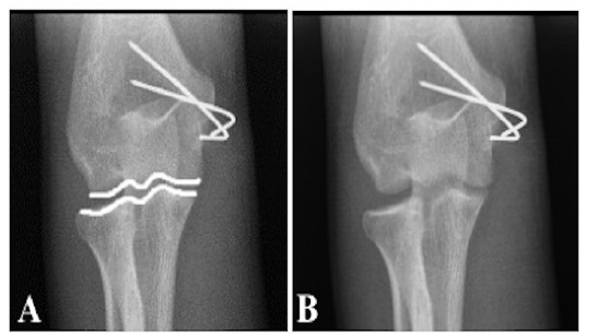
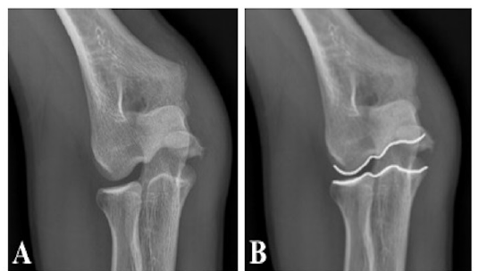
جميع المرضى لديهم علامات طبية وإشعاعية للشفاء في المتابعة النهائية There was no radiographic evidence of loss of reduction at intervals or at the final follow-up. There were no cases of remaining deformity or instability. لا أحد من المرضى كان لديه أعراض عصبية قبل التشغيل وقد تم تحديد المعيار التشخيصي الجديد للإشعاع في جميع مرضى الكسور الوسيطين المحتجزين.
المناقشة
The mechanism of injury for medial epicondyle fractures of the distal humerus has been suggested to be a fall on an outstretched arm with the elbow in extension and the wrist in supination and extension, which forces the elbow into valgus stress. القوة الناجمة عن ذلك تؤدي إلى إزدهار وسيطة (6, 7)تم الإبلاغ عن وجود حوالي 60 في المائة من حالات الكسور الوسيطة المرتبطة بتفكك القوس (8)The medial epicondyle is incarcerated in the joint in 15% to 25% of cases (5, 9)في بعض الأحيان قد ينتقل القوس تلقائياً وأشعة الضغط على الوادي قد تساعد في تشخيص الإصابات المصاحبة (8, 9).
ومعاملة الكسور الوسيطة في الهضم أمر مثير للجدل. غير أن النهج الجراحي يشار إليه بوضوح في حالات الاصطدام الداخلي للجزء، أو للاشتباه في اغتصاب الأعصاب الحلقية، أو في حالات عدم الاستقرار الملحوظ للقوس (10)وقد أفيد في المؤلفات أن معدلات النقابة أعلى من الكسور الوسيطة المثبتة جراحيا بالمقارنة مع الكسور المعالجة بتحفظ. غير أنه في تحليل مميت بواسطة كاماث وآخرون، لم يتم تحديد أي فرق كبير في النتيجة الوظيفية بين المرضى الذين يتلقون العلاج الجراحي أو المحافظ. ومن المثير للاهتمام أن المرضى الذين لا يتلقون العلاج الجراحي يقل ألمهم على الرغم من انخفاض معدلات النقابة. ومن ثم، فقد تم قبول أن اتحاد الوسطاء لا يرتبط بالنتائج الوظيفية (11)وعلى الرغم من ملاحظة الشفاء الإشعاعي في جميع المرضى في الدراسة الجارية في إطار المتابعة النهائية، فإن هذا لم يكن هدف العلاج. In this study, good outcomes were achieved with operative treatment for incarcerated medial epicondyle fractures of the distal humerus. وكانت نتائج الدراسة قابلة للمقارنة مع المؤلفات الحالية (9, 12, 13).
وقد سُجلت تدخلات جراحية متعددة في الأدبيات مثل إصلاح الخياطة، وتثبيت أسلاك كيرشنر، وتثبيت الشظايا ومسح الأنسجة الناعمة لوسائط الإعلام (3–5, 14)- وفي الدراسة الحالية، استخدمت أسلاك كيرشنر للتثبيت، ووجد أن هذا أسلوب فعال للتثبيت. وقد ذكر المؤلفون الذين يدعون إلى إصلاح المسامير كطريقة التثبيت أنه مع إصلاح المسامير يمكن تطبيق التعبئة المبكرة بعد التشغيل. However, they also used a splint or cast for two weeks after the operation to protect the fracture displacement (1)- إن سلك كيرشنر سهل وأقل تعقيدا نسبيا، ولا توجد مضاعفات تتعلق بطريقة التثبيت التي وضعت في الدراسة الحالية.
Surgically treated medial epicondyle fractures have been reported to betrophic, with a deformity of the medial epicondyle and with an “ulnar sulcus,” in many cases (15)- لم يكن لدى أي من المرضى في الدراسة الحالية تشوهات في الكبريت في نهاية فترة المتابعة. وعلى الرغم من صعوبة تشخيص الكسور الوبائية الوسيطة عندما يغيب التشخيص، فقد أُبلغ عن حالات تأخير في الاضطرابات العصبية الرئوية في الأدبيات (10, 16, 17)- يمكن أن يؤدي التشخيص المفقود للاختطاف داخل الأعياد في الإسطوانات الوسيطة إلى فرض قيود كبيرة على التنقل وزيادة خطر الإصابة بالمرض العصبي الرئوي. والتدخل المبكر ضروري لأن التشخيص يزداد سوءا مع مرور الوقت، نظرا لضعف التعافي من القيود على الأعصاب الناجمة عن الكبريت.
قد تغيب عن الأشعة التقليدية بسبب صغر حجم الشظية الكسورية الوسيطة المرتبطة بتفكك النوافذ في الأطفال، ويمكن أن يُخطئ في مركز التجميل، وقد يُخفى الموقف وراء الهرطبة الفاشلة (10)- إذا كان الفطريات المنبعثة تبدو غير متماثلة على جهاز لاسلكي داخلي، أو إذا كانت الصرعات مرئية على الراديو الجانبي، فلا بد من تشريد الكوكونيل (18, 19)كما أن الكسور الوسيطة المسجونة بعد تفكك النواة في الأطفال هي كيان نادر، ولكن بسبب ما يرتبط بذلك من خطر الإصابة بالأعصاب الرئوية والتقييد الجسيم للحركة النبيلية، فإن التشخيص الفوري لهذا الكسر وعلاجه أمر بالغ الأهمية. ونظرا للصعوبات التي واجهتها خلال تشخيص هذه الكسور، حددنا علامة إشعاعية جديدة يمكن أن يلاحظها بسهولة جراحو العظام بعد تشتيت القوس في الأطفال. The weak point of the study was the small number of cases and that the study was retrospective.
The results of this study suggest that open reduction internal fixation of the incarcecerated epicondyle fractures after elbow dislocation resulted in a satisfactory motion and function. In the pediatric population, fracture may not easily diagnosed on conventional radiographs. وهذا المعيار التشخيصي الإشعاعي الجديد الذي حددته هذه الدراسة مفيد، ولا يؤدي إلى أي تكاليف إضافية، وقد يحل مشاكل التشخيص التي يعاني منها الكسور الوسيطة المتسربة في الرطوبة بعد تفكك القاع في السكان الأطفال.
الإقرارات
- مساهمات المؤلفين (CRediT)
- ساهم جميع المؤلفين بالتساوي في جميع أدوار CRediT الأربعة عشر (Conceptualization, Data curation, Formal analysis, Funding acquisition, Investigation, Methodology, Project administration, Resources, Software, Supervision, Validation, Visualization, Writing – original draft, Writing – review & editing).
- التمويل
-
Funding information was not reported in the published version of this article.
- تضارب المصالح
- The authors declare that they have no conflict of interest in the current study.
- الموافقة الأخلاقية
- The study was approved by the Institutional Review Board.
- إتاحة البيانات
- A data availability statement was not reported in the published version of this article.
المراجع (19)
13 من أصل 19 مرجعًا مرتبطة بمعرّف DOI.
- Gottschalk HP, Eisner E, Hosalkar HS. Medial epicondyle fractures in the pediatric population. J Am Acad Orthop Surg 2012;20:223– 32. DOI Crossref PubMed Google Scholar
- Kobayashi Y, Oka Y, Ikeda M, Munesada S. Avulsion fracture of the medial and lateral epicondyles of the humerus. J Shoulder Elbow Surg 2000;9(1):59-64. DOI Crossref PubMed Google Scholar
- Fowles JV, Slimane N, Kassab MT. Elbow dislocation with avulsion of the medial hu meral epicondyle. J Bone Joint Surg Br 1990;72(1):102-4. DOI Crossref PubMed Google Scholar
- Farsetti P, Potenza V, Caterini R, Ippolito E. Long-term results of treatment of frac tures of the medial humeral epicondyle in children. J Bone Joint Surg Am 2001;83(9):1299-305. DOI Crossref PubMed Google Scholar
- Wilson NI, Ingram R, Rymaszewski L, Miller JH. Treatment of fractures of the medial epicondyle of the humerus. Injury 1988;19(5):342-4. DOI Crossref PubMed Google Scholar
- Beaty JH, Kasser JR (2010) The elbow: physeal fractures, apophyseal injuries of the distal humerus, osteonecrosis of the trochlea, and T-condylar fractures. In: Beaty JH, Kasser JR (eds) Rockwood and Wilkins’ fractures in children, 7t h ed. Lippincott Williams & Wilkins, Philadelphia, pp 566–77. PubMed Google Scholar
- Smith BG, Pierz KA (2011) Open reduction and internal fixation of fractures of the me dial epicondyle. In: Flynn JM, Wiesel SW (eds) Operative techniques in orthop edic surgery, vol:2. Lippincott Williams&Wilkins, Philadelphia, pp 1042–5. PubMed Google Scholar
- Lindham S, Hugosson C. The significance of associated lesions including dislocation in fractures of the neck of the radius in children. Acta Orthop Scand 1979;50(1):79-83. DOI Crossref PubMed Google Scholar
- Lee HH, Shen HC, Chang JH, Lee CH, Wu SS. Operative treatment of displaced medial epicondyle fractures in children and adolescents. J Shoulder Elbow Surg 2005;14 (2):178-85. DOI Crossref PubMed Google Scholar
- Lima S, Correia JF, Ribeiro RP, Martins RM, Alegrete N, Coutinho J, Costa GM. A rare case of elbow dislocation associated with unrecognized fracture of medial epicondyle and delayed ulnar neuropathy in pediatric age. J Shoulder Elbow Surg 2013;22(3):e9–e11. DOI Crossref PubMed Google Scholar
- Kamath AF, Baldwin K, Horneff J, Hosalkar HS. Operative versus nonoperative management of pediatric medial epicondyle fractures: A systematic review. J Child Orthop 2009;3(5):345-57. DOI Crossref PubMed Google Scholar
- Ip D, Tsang WL. Medial humeral epicondylar fracture in children and adolescents. J Orthop Surg (Hong Kong) 2007;15(2):170-3. DOI Crossref PubMed Google Scholar
- Hines RF, Herndon WA, Evans JP. Operative treatment of medial epicondyle fractures in children. Clin Orthop Relat Res 1987;223:170-4. DOI Crossref PubMed Google Scholar
- Gilchrist AD, McKee MD. Valgus instability of the elbow due to medial epicondyle nonunion: Treatment by fragment excision and ligament repair. A report of 5 cases. J Shoulder Elbow Surg 2002;11(5):493-7. DOI Crossref PubMed Google Scholar
- Skak SV, Grossmann E, Wagn P. Deformity after internal fixation of fracture separation of the medial epicondyle of the humerus. J Bone Joint Surg Br 1994;76(2):297-302. DOI Crossref PubMed Google Scholar
- Haflah NHM, Ibrahim S, Sapuan J, Abdullah S. An elbow dislocationin a child with missed medial epicondyle fracture and late ulnar nerve palsy. J Pediatr Orthop B 2010;19:459-61. PubMed Google Scholar
- Rasool MN. Dislocation of the elbow in children. J Bone Joint Surg Br 2004;86- B:1050-8. PubMed Google Scholar
- Patrick J. Fracture of the medial epicondyle with displacement into the elbow joint. J Bone Joint Surg 1946;28:143-7. PubMed Google Scholar
- Martini M, Hallaj N, Daoud A, Descamps L. Les luxations traumatiques recentes du coude: a propos de 94 observations. Acta Orthop Belg 1978;44:542-54. PubMed Google Scholar
اقرأ النص الكامل
افتح ملف PDF هنا
جارٍ تحميل ملف PDF…
إذا لم يظهر ملف PDF هنا، استخدم زر التنزيل أعلاه.
كيف تقتبس هذه المقالة
Ozdemir G, Deveci A, Bingol O, Kilic E, Ceyhan E, Turan S. A Blocking Barrier During Reduction of the Elbow Dislocation in Children: Incarcerated Medial Epicondyle Fracture. Ulutas Med J. 2018;4(2):103-107. doi:10.5455/umj.20171107113200
Ozdemir, G., Deveci, A., Bingol, O., Kilic, E., Ceyhan, E., & Turan, S. (2018). A Blocking Barrier During Reduction of the Elbow Dislocation in Children: Incarcerated Medial Epicondyle Fracture. The Ulutas Medical Journal, 4(2), 103–107. https://doi.org/10.5455/umj.20171107113200
Ozdemir, G. et al. (2018) 'A Blocking Barrier During Reduction of the Elbow Dislocation in Children: Incarcerated Medial Epicondyle Fracture', The Ulutas Medical Journal, 4(2), pp. 103–107. doi: 10.5455/umj.20171107113200.