The Ulutas Medical Journal · ISSN 2149-0430 (print), 2149-388X (online) · https://www.ulutasmedicaljournal.com/?mno=16164
Original Article · Open access
The Use of Fibrin Sealant in Chiari Malformation Type-1 Surgery; Is It Effective in Preventing the Formation of CSF Fistula?
- Department of Neurosurgery, Medicine Hospital, Biruni University, Istanbul, Turkey ROR
- Department of Pediatrics, Medicine Hospital, Biruni University, Istanbul, Turkey ROR
Abstract
- Background
- Symptomatic disease requires surgical intervention comprising of posterior fossa decompression with or without an expansile duraplasty. Until date, the optimal surgical treatment for CM-1 has not been delineated. Thus, we evaluated the results to assess the effectiveness of fibrin sealant augmentation in combination with locally harvested autologous pericranium for duraplasty in adult CM-1 decompression.
- Materials and Methods
- We retrospectively evaluated the cases that were operated due to CM-1. The cases were divided into two groups in which fibrin sealant (FS) was used and not used. There were 15 patients in the first group (FS group) and 16 in the second group (no fibrin sealant (NFS) group). We followed the standardized surgical technique suggested by Stevens and colleagues for CM 1 decompression.
- Results
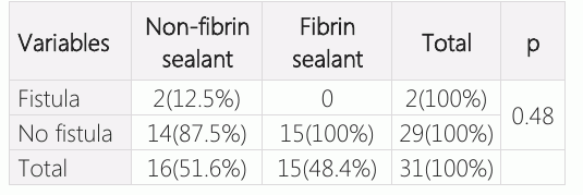
- Cerebrospinal fluid (CSF) fistula was not observed in any patient in the FS group. However, in the NFS group, two patients were reported to develop CSF fistula. Although the findings are not statistically significant, these are clinically important.
- Conclusion
- Use of FS to seal the autologous graft during closure may prevent the occurrence of CSF fistula among patients of CM-1.
Keywords
Full text
Introduction
Chiari malformation has been named after an Austrian pathologist Hans Chiari, who recognized this disorder in 1890s. There are many synonyms of CMlikehindbrain herniation, Arnold-Chiari malformation and tonsillar ectopia. It’s of five types ranging from Type 0 to 4. Of these, CM-1 is the most common type, which involves the lower part of the cerebellum known as tonsils. However, it does not involve brain stem. The symptoms of CM-1 commonly appear in second decade of life that is in adult hood. This is because CM-1 develops when the skull and brain are in the growing stage, so it goes unnoticeable in infancy and childhood (1–3). The diagnosis of this disorder in most of the cases is incidental. That too has become possible due to availability of more advanced and sophisticated diagnostic techniques like Magnetic Resonance Imaging (MRI). The common symptoms appearing in the patients with this disorder are headache at the back of head and nape of neck, dizziness, numbness or tingling sensations in hands and feet, difficulty in walking due to improper balance, loss of pain or temperature sensations of upper body in few cases etc. These all symptoms are attributed to functions coordinated by cerebellum like balance, vision, speech and coordination. Patients with CM 1 often present with other neurological abnormalities like syringomyelia, hydrocephalus etc. (1–3).
The treatment of CM 1 depends upon severity and symptoms of disease. Surgery is the best resort to minimize the existing symptoms. Though the surgery can’t provide the reversal of nerve damage, but it prevents further damage and limit the symptoms. However, there are few concerns regarding the surgery which still needs to be addressed like how much should be the extent of bony removal, size of dural opening, necessity of expanding dural space, material to be used for duraplasty and whether augmentation with FS is required to prevent further complications or not. These issues are still controversial and needs answer to delineate the surgical intervention. There is insufficient literature on the efficacy of fibrin sealant usage for dura closure, hence we investigated the effect of fibrin sealant usage on the results. Therefore, this analysis was performed to evaluate the use of fibrin sealant in CM-1 decompression to prevent postoperative CSF fistula development in adults.
Material and Methods
In clinical retrospective analysis approved by the local ethics committee, we evaluated cases operated for CM-1 from 2009 to 2018. The cases were divided into two groups in which fibrin sealant (FS) was used and not used. There were 15 patients in FS group and 16 patients in non-fibrin sealant (NFS) group.
The patients were evaluated according to age, gender, presenting symptoms and additional pathologylikesyringomyelia, findings of MRI for tonsillar herniation, whether FS was used, and fistula developed.
Surgical Procedure
Patients were positioned prone on the operative table. Pin head holder was not used in majority of the cases. A midline incision was made one inch above the inion extending inferiorly to the level of mid cervical spine. We extended the incision superiorly and performed a blunt dissection of the subgaleal connective tissue. This helped to generate adequate amounts of pericranium for dural grafting. Posterior fossa and C1 decompression were performed. The dimensions of posterior fossa craniectomy and C1 laminectomy were 3.5-4 *3.5-4 cm and 1.5-2 cm respectively. The dura was opened sharply in a Y-shaped fashion exposing the inferior aspect of the cerebellar hemispheres. No tonsillar resection was done. We did not use soft tissue repair patch also. A thin layer of FS was applied over the suture line and the entire graft before closure in 15 patients whereas FS was not applied in 16 patients. The surgical wound was closed in multiple layers beginning with muscle, ligamentum nuchae, subcutaneous tissue, and then, skin. Patients were discharged from hospital and were followed up to assess if they developed CSF fistula or not.
Statistical Analysis
Descriptive analysis was used to describe the data in terms of age, gender, MRI findings, associated symptoms. Chi-square test was used to assess the efficacy of FS to prevent CSF fistula among FS group and NFS group.
Results
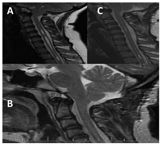
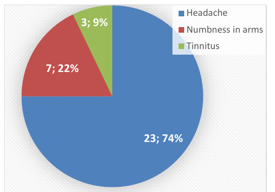
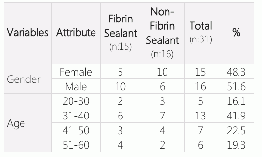
31 patients were included in the study, the overall range of age varied from 23 years to 59 years with the median age of 40 years. Majority of the patients were in the age group of 31 to 40 years (n:13) followed by 7 patients in the age group of 41- 50 years and six patients were aged more than 50 years. There were only five patients in the age group of 20-30 years. There were 15 women and 16 men in total. Table-1 describes the baseline characteristics of groups. The patients presented with varied symptoms. The most common ones were headache (74% cases) followed by numbness or pain in the arms (22%) and rest had tinnitus as the main symptom (Figure-2). There were eight individuals who had multiple combination of symptoms. MRI of the patients showed that tonsil herniation ranged from 7 mm to 29 mm (median: 13 mm) (Figure-1). Syringomyelia was found in nine cases. Of these 31 patients, in 15 patient’s dura closure was done with FS and no CSF fistula developed. In other group, where FS was not used, CSF fistula occurred in two cases. Patients with CSF fistula were treated with lumbar puncture and primary suture. There is no statistically difference in the incidence of CSF fistula among patients who underwent CM-1 decompression by using FS and those patients in whom FS was not used during duraplasty (p=0.48)(Table-1 and 2).
Discussion
In a review, it was concluded that indication for surgery among patients with CM 1 depends upon the age, severity of symptoms and presence of brainstem dysfunction, cranial nerve dysfunction and syringomyelia. These complications mandate surgical intervention to prevent further adverse outcomes. The asymptomatic patients with CM 1 are generally not considered eligible to undergo surgery (22). In this study also, for surgery we recruited patients who had severe presenting symptoms. Eligibility of patients for surgical intervention was further strengthened by MRI findings regarding the extent of tonsillar herniation and presence of complications like syringomyelia. From literature review, it could be concluded that decision to do surgery or not for correcting tonsils in CM-1 is still a controversial issue, despite the better understanding of the patho physiological mechanisms and all the recent advances in imaging.
Surgical intervention in the treatment of symptomatic CM 1 cases has been shown to be needed, moreover, recent studies show that it would be better to perform treatment without progress. It implies that even if the symptoms are not that serious to take action, but it is good to undergo elective surgery to prevent future complications (2–6). Once the decision is made to conduct surgical intervention, there are numerous surgical approaches available for the treatment of these patients (2–4, 6–10). There is no concluding evidence which approach is better over other. However, the most common approach followed by neurosurgeons for treating adult patients is the one we have used in our cases which was described by Stevens et al (23).
Most of the published case series and case reports advocate that suboccipital craniectomy, removal of the posterior arch of C1, and an augmentative duraplasty represent the best baseline surgical approach which can be used with slight modifications to suit the need of individual patient.Suboccipital posterior fossa decompression and C1 laminectomy are done as standardized procedures with a success rate of 95% to 97%. (3, 7, 11–13).
In our cases, a similar approach was followed to treat patients. In addition to this, performance of an expandable and augmentative duraplasty for avoiding any postoperative compression of the posterior fossa contents seems to be widely accepted especially among adults (3, 7, 14, 15). The application of this approach among pediatric patients is still controversial. Another related debatable issue which made us to conduct this research pertains to which material should be used for performing the duraplasty? The use of both autograft and allograft has been advocated in the literature. Mottolese et al. (8) reported that the usage of soft tissue repair patch along with a FS resulted in better outcome rates in their series. However, the authors noticed an increased incidence of complications in those cases that a dural allograft and a FS were utilized. Hence, we used autograft, which does not trigger immune response, instead of soft tissue repair patch to prevent development of complications like aseptic meningitis.
Surgical intervention is associated with a wide spectrum of intraoperative and postoperative complications. One such common complication is the postoperative development of a CSF leakage, independent of the type of the employed surgical approach. While using dural grafts, holes are produced by surgical needles which lead to CSF leaks. (2) Hence, FS have been developed to prevent complications like CSF leaks, low-pressure headaches and pseudo meningoceles. The incidence of CSF leak has been reported to be as high as 28% in p-fossa procedures as it is located at the dependent portion of the skull base (16). Till date, studies have been published using FS alone or in combination with autologous duraplasty materials in cranial surgeries (17–21). One study reported 4.5% incidence of CSF leakage and an infection rate of 8.1% (17). According to another study, a polyethylene glycol (PEG) hydrogel sealant was used and no CSF leak was reported after three months of follow-up (18, 19). There were no CSF leaks in our FS where we used FS. However, in NFS group where we did not use FS, there were two cases who developed CSF fistula. Although the findings are not statistically significant but clinically, it strengthens the literature for the use of a FS in combination with autologous pericranium for dural closure. The reason for insignificant findings could be due to the small number of cases. Hence, in this case, clinical relevance of effectiveness is more important as compared to statistical significance. We retrospectively reviewed the results of the patients. We carried out surgeries with the standardized method along with usage of autologous graft. Therefore, although this study is inadequate to reach important findings, it is methodologically robust. Studies with bigger number of cases should be conducted to address all the identified controversies in the literature. Studies on a greater number of patients are required to provide better objective results.
Conclusion
Clinically, use of FS for duraplasty prevented the development of the CSF fistula in CM-1 patients compared to the group in which the CSF fistula developed in 2 cases was not used. Use of FS to seal the autologous graft during closure may prevent occurrence of CSF fistula in patients.
Declarations
- Author contributions (CRediT)
- All authors contributed equally to all 14 CRediT roles (Conceptualization, Data curation, Formal analysis, Funding acquisition, Investigation, Methodology, Project administration, Resources, Software, Supervision, Validation, Visualization, Writing – original draft, Writing – review & editing).
- Funding
-
Funding information was not reported in the published version of this article.
- Conflicts of interest
- None of the authors has a conflict of interest with the submission.
- Ethics approval
- In clinical retrospective analysis approved by the local ethics committee, we evaluated cases operated for CM-1 from 2009 to 2018. The cases were divided into two groups in which fibrin sealant (FS) was used and not used.
- Data availability
- A data availability statement was not reported in the published version of this article.
References (23)
16 of 23 references are linked to their DOI.
- D Adams CB. I’ve torn it: How to repair it. Br J Neurosurg 1995; 9:201– 4 DOI Crossref PubMed Google Scholar
- Haines SJ, Berger M. Current treatment of Chiari malformations types I and II: a survey of the pediatric section of the American association of Neurological Surgeons. Neurosurgery 1991:28(3):353-57 DOI Crossref PubMed Google Scholar
- Nash J, Cheng JS, Meyer GA, Remler BF. Chiari type I malformation: overview of diagnosis and treatment. Wisconsin Medical Journal 2002;101(8):35-40 DOI Crossref PubMed Google Scholar
- Nagib MG. An approach to symptomatic children (ages 4 – 14 years) with Chiari type-I malformation. Pediatr Neurosurg 1994;21(1):31-35 PubMed Google Scholar
- Dyste GN, Menezes AH. Presentation and management of pediatric Chiari malformations without myelodysplasia. Neurosurgery 1998;23(5):589-97 PubMed Google Scholar
- Krieger MD, McComb JG, Levy ML. Toward a simpler surgical management of Chiari I malformation in pediatric population. Pediatr Neurosurg 1999;30(3);113-21. DOI Crossref PubMed Google Scholar
- Zerah M. Syringomyelia in children. Neurochirurgie 1999; 1(45):37-57 PubMed Google Scholar
- Lazareff JA, Valencia-Myoral P. Chiari malformation. Boletín Médico del Hospital Infantil de México 1990;47 (10):719-24 PubMed Google Scholar
- Eule Jm, Erickson MA, O’Brien MF, Handler M. Chiari I malformation associated with syringomyelia and scoliosis: a twenty-year review of surgical and nonsurgical treatment in a pediatric population. Spine 2002;27(13): 1451-55 DOI Crossref PubMed Google Scholar
- Yundt KD, Park TS, Tantuwaya VS, Kaufman BA. Posterior fossa decompression without duraplasty in infants and young children for treatment of chiari malformation and achondroplasia. Pediatric Neurosurgery 1996;25(5):221– PubMed Google Scholar
- Isu T, Sasaki H, Takamura H, Kobayashi N. Foramen magnum decompression with removal of the outer layer of the dura as treatment for syringomyelia occurring with Chiari I malformation. Neurosurgery 1993;33(5): 844–849 DOI Crossref PubMed Google Scholar
- Genitore L, Peretta P, Nurisso C, Macinante L, Mussa F. Chiari type I anomalies in children and adolescents: minimally invasive management in a series of 53 cases. Child's Nervous System 2000;16(10-11):707–718 PubMed Google Scholar
- Alden TD, Ojeman JG, Park TS. Surgical treatment of Chiari malformation: indications and approaches. Neurosurgical Focus;2001;11(1):E2 DOI Crossref PubMed Google Scholar
- Hankinson T, Tubbs RS, Wellons JC. Duraplasty or not? An evidence-based review of pediatric Chiari malformation. Child's Nervous System 2011; 27(1):35– 40 DOI Crossref PubMed Google Scholar
- Sindou M, Gimbert E. Decompression for Chiari type I-malformation (with or without syringomyelia) by extreme lateral foramen magnum opening and expansile duraplasty with arachnoid preservation: comparison with other technical modalities (Literature review). Advances and Technical Standards in Neurosurgery 2009;34:85-110 DOI Crossref PubMed Google Scholar
- Manley GT, Dillon W. Acute posterior fossa syndrome following lumbar drainage for treatment of suboccipital pseudomeningocele. Report of three cases. J Neurosurg. 2000; 92:469– 74 DOI Crossref PubMed Google Scholar
- Summary of Safety and Effectiveness Data: Dura Seal Dural Sealant System. Silver Spring: U.S. Food and Drug Administration; 2005. Administration USFaD PubMed Google Scholar
- Cosgrove GR, Delashaw JB, Grotenhuis JA, Tew JM, Van Loveren H, Spetzler RF, et al. Safety and efficacy of a novel polyethylene glycol hydrogel sealant for watertight dural repair. J Neurosurg. 2007; 106:52– 8 DOI Crossref PubMed Google Scholar
- Than KD, Baird CJ, Olivi A. Polyethylene glycol hydrogel dural sealant may reduce incisional cerebrospinal fluid leak after posterior fossa surgery. Neurosurgery 2008 Jul;63: ONS182-6; discussion ONS186-7 DOI Crossref PubMed Google Scholar
- Lam FC, Penumaka A, Chen CC, Fischer EG, Kasper EM. Fibrin sealant augmentation with autologous pericranium for duraplasty after suboccipital decompression in Chiari 1 patients: A case series. Surg Neurol Int. 2013; 4:6 DOI Crossref PubMed Google Scholar
- Tonkins M, Farooqi N, Ahmed R, Sinha S, Bhattacharyya D. Closing the dura: dural hitching versus surgicel and tisseel overlay graft in craniocervical decompression for Chiari 1 malformation. Br J Neurosurg. 2017 Aug;31(4): 422-425 DOI Crossref PubMed Google Scholar
- Langridge B, Phillips E, Choi D. Chiari Malformation Type 1: Systematic Review of Natural History and Conservative Management. World Neurosurg. 2017 Aug;104:213-219 DOI Crossref PubMed Google Scholar
- Stevens EA, Powers AK, Sweasey TA, et al. Simplified harvest of autologous pericranium for duraplasty in Chiari malformation Type I. Technical note. J Neurosurg Spine 2009;11:80-3 DOI Crossref PubMed Google Scholar
Read the full text
Open the PDF here
Loading the PDF…
If the PDF does not show here, use the download button above.
How to cite this article
Dogu H, Ozdemir AA. The Use of Fibrin Sealant in Chiari Malformation Type-1 Surgery; Is It Effective in Preventing the Formation of CSF Fistula?. Ulutas Med J. 2019;5(1):65-70. doi:10.5455/umj.20191108075317
Dogu, H., & Ozdemir, A. A. (2019). The Use of Fibrin Sealant in Chiari Malformation Type-1 Surgery; Is It Effective in Preventing the Formation of CSF Fistula?. The Ulutas Medical Journal, 5(1), 65–70. https://doi.org/10.5455/umj.20191108075317
Dogu, H. and Ozdemir, A.A. (2019) 'The Use of Fibrin Sealant in Chiari Malformation Type-1 Surgery; Is It Effective in Preventing the Formation of CSF Fistula?', The Ulutas Medical Journal, 5(1), pp. 65–70. doi: 10.5455/umj.20191108075317.