The Ulutas Medical Journal · ISSN 2149-0430 (印刷版), 2149-388X (在线版) · https://www.ulutasmedicaljournal.com/?mno=87160
病例报告 · 开放获取
Colon Cancer Micking Cholangiocarcinoma和化学疗法诱导的胆碱酯酶:案例报告
标题、摘要和全文为机器翻译。 英文原文
- Department of Radiology, Chi Mei Medical Center, Liouying, Tainan, Taiwan ROR
- Department of Intensive Care Medicine, Chi Mei Medical Center, Liouying, Tainan, Taiwan ROR
- Department of Nursing, Kaohsiung Municipal United Hospital, Taiwan ROR
- 开放获取
- CC BY-NC 4.0
- ORCID
- 资助
- 伦理批准
- 数据可用性
摘要
在没有肝癌组织参与的情况下,肠癌的内侧元化非常罕见,可造成双胞胎障碍。 我们在此介绍一个罕见的结肠癌变异症病例,该病是在右侧肝电解后,在接受化疗期间发展而来的。 腹部和腹部直肠切除术和排水术(PTCD)的计算成形图(CT)是因为阻塞性黄油。 CT导体活检显示,高级非典型腺体是CK7(â),CK20(+),和CDX-2(+);元静脉动腺瘤将首先被考虑,而不是胆固醇癌。 临床医生应意识到,结肠癌的细胞内元化可以模仿胆固醇癌和化疗引起的结肠炎.
关键词
全文
全文为机器翻译;以英文原文为准。 英文原文
导言
在没有肝癌参与的情况下,结肠癌的内侧元化是罕见的 (1)肝脏的元素可造成双胞胎阻塞,并导致无痛的黄undi,苍白的凳子,暗的尿液和棱柱炎,这种元素与直接双胞胎和碱性磷脂酶的升高有关. (2)此外,图像发现显示,仿制多焦性双管紧固性结膜炎等元瘤是罕见的。 这里,我们报告一个结肠癌的细胞内元化病例 以及相关的成像结果。
案例介绍
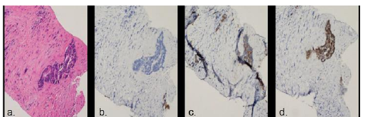
一名82岁男子在接受化疗期间,在右侧肝电解后,呈上结肠元化。 定期后续的实验室测试显示CA19-9和CEA升高. 进行了计算成的直肠造影术(CT),揭示了普通胆管(CBD)和右肝内膜(IHD)的软组织密度,并加厚了墙壁,导致双胞胎对映(图1和图2)。 进行了直肠切除术和排水术(PTCD),切除术揭示出肝内和外肠管的多种严格性,造成阻塞性黄油(图3)。 根据他的病史和放射性发现,不同的诊断包括元化、化疗引起的胆固醇炎(CISC)和胆固醇癌。 从直肠截流排水管中采集的血液的细胞学没有发现恶性特征。 随后进行了CT导体活检,并揭示出高级非典型腺体. 免疫组织化学研究表明,非典型腺体是CK7(−),CK20(+),和CDX-2(+)(图4).
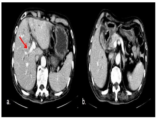
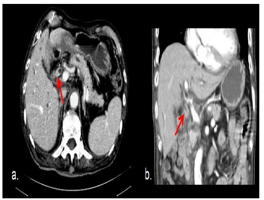
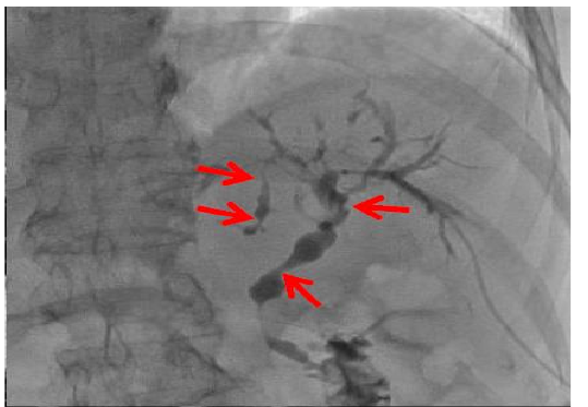
根据研究结果,由于没有CK7的表达,将首先认为是元静脉瘤,而不是双管主要恶性。 病人的病情不佳,总的比里鲁宾水平再次上升,肝功能恶化,需要每侧插入两个PTCD. 一个月后,化脓性硅道感染逐渐恶化,患者最终去世.
讨论情况
在我们的案例中,CT和乔兰吉格文学显示,外肝和肝内胆管的墙壁加厚,并伴有严格和分化。 我们的病例还提供了子宫内膜阻塞和子宫内膜收缩的成像结果,这些也是中继血球结膜炎和初级血球癌的成像结果。 据报道,CISC偶尔是由肝动脉输液化疗与氟化py引起的. (3–5)在研究中,考虑到化疗(FOLFOX4)中突然爆发胆汁胶囊炎(FOLFOX4),疑似胆囊炎,患者初步接受了类固醇治疗,化疗也停止了. 然而,快速升高的肿瘤标记(CEA:171纳克/mL,CA19-9:10115 U/mL),在停止化疗后没有出现改善. 诸如“洋葱皮肤”纤维化等结膜炎的区别发现 (6) 在我们的肝脏活检中没有发现 Cholangiocarcinoma的形态和生长形态如下: 质地形成,地层渗透,以及地层内生长. (7)细胞内和腹膜渗入型胆固醇癌的成像结果包括:脊髓收缩、细胞内肿瘤、有或无增强的焦或弥散性胶质增厚。 (8, 9)与主要肝内胆碱性胆碱酯酶瘤区别开来是困难的。 然而,免疫组织化学研究,如CK7,CK20和CDX2污迹研究,可以提供明确的诊断. (10). Metastatic crooma 显示 CK20 阴性与 CK7 阴性,与相邻的 bile 管道上皮形成对比。 (11) 。在我们的情况下,没有找到 CK7 表达式。 根据结果,元静脉瘤将被视为第一,而不是初级双管恶性。 在我们的案例中, 成像研究 发现阻塞性黄蜂 没有间隙质量。 腹腔内损伤的生物检查技术包括:刷子细胞学、胆囊镜下蛤壳强力和氟镜下蛤壳强力。 在前一次研究中,胆汁镜定向活检技术具有最高的敏感性和特异性,但这种差异在统计学上并不显著. (12)然而,在这项研究中,病人拒绝接受内分泌的复方胆囊造影术(ERCP),而我们的医院没有蛤壳硬体造影术;因此,我们进行了跨肝活体活体检查。 由于在声学下无法很好地识别软组织密度的胶带增厚,所以实时USG与CT结合对于将不显眼的焦肝损伤本地化很有用. (13)肝元化的细胞内生长的病原学不详,但病原学可能涉及血管入侵或通过胆囊流将恶性细胞植入细胞内. (14)体内元病患者的预后比无元病患者要好;此外,有宏观参与的患者的5年存活率(80%)远高于有微观参与的患者. (15)然而,我们病人的病情很差,他的总比里鲁宾水平再次上升,他的肝功能恶化,甚至在两侧插入了两颗三联疫苗之后;因此,病人被转移到了收容所,一个月后死亡。
结论
最后,很容易忽略了生物内元病。 免疫组织化学研究有助于实现正确的诊断和管理.
声明
- 作者贡献(CRediT)
- 所有作者对全部 14 项 CRediT 角色贡献均等(Conceptualization, Data curation, Formal analysis, Funding acquisition, Investigation, Methodology, Project administration, Resources, Software, Supervision, Validation, Visualization, Writing – original draft, Writing – review & editing)。
- 资助
-
Funding information was not reported in the published version of this article.
- 利益冲突
- All authors declare that there is no conflict of interest.
- 伦理批准
- Ethics approval details were not reported in the published version of this article.
- 数据可用性
- A data availability statement was not reported in the published version of this article.
参考文献 (15)
15 条参考文献中有 13 条已链接 DOI。
- Patel T. Increasing incidence and mortality of primary intrahepatic cholangiocarcinoma in the United States. Hepatology. 2001;33:1353–1357 DOI Crossref PubMed Google Scholar
- Nagler J, Rochwarger AM. Metastatic colon carcinoma simulating primary bile duct carcinoma via endoscopic cholangiography. Gastrointest Radiol. 1977;2:75–76 DOI Crossref PubMed Google Scholar
- Sandrasegaran K, Alazmi WM, Tann M, et al. Chemo therapy-induced sclerosing cholangitis. Clin Radiol. 2006;61:670–8 PubMed Google Scholar
- Shea WJ Jr, Demas BE, Goldberg HI, et al. Sclerosing cholangitis associated with hepatic arterial FUDR chemotherapy: Radiographic-histologic correlation. AJR Am J Roentgenol. 1986; 146:717–21 DOI Crossref PubMed Google Scholar
- Kemeny N, Daly J, Oderman P, et al. Hepatic artery pump infusion: Toxicity and results in patients with metastatic colorectal carcinoma. J Clin Oncol. 1984; 2:595-600 85 77 Intrabiliary Metastasis of Colon Cancer Guang-Mao Lee et al. DOI Crossref PubMed Google Scholar
- Akira K, Kazuyoshi O, Nobuyasu F, et al. Chemo therapy-Induced Sclerosing Cholangitis Caused by Systemic Chemotherapy, ACG Case Rep J 2019;6:1-4 PubMed Google Scholar
- Yamasaki S. Intrahepatic cholangiocarcinoma: macro scopic type and stage classification. J Hepato biliary Pancreat Surg 2003; 10:288-291 DOI Crossref PubMed Google Scholar
- Lee WJ, Lim HK, Jang KM, et al. Radiologic spectrum of cholangiocarcinoma: emphasis on manifestations and differential diagnoses. Radio Graphics 2001;21(spec issue):S97–S116 DOI Crossref PubMed Google Scholar
- Han JK, Choi BI, Kim AY, et al. Cholangiocarcinoma: pictorial essay of CT and cholangiographic findings. RadioGraphics 2002;22:173–187 DOI Crossref PubMed Google Scholar
- Dong Y, Patel H, Patel C. Intrabiliary hepatic metastasis of colorectal carcinoma mimicking primary cholangio carcinoma: a case report and review of the literature. Case Rep Pathol. 2016; 4704781 DOI Crossref PubMed Google Scholar
- Tokai H, Kawashita Y, Eguchi S, et al. A case of mucin producing liver metastases with intrabiliary extension. World J Gastroenterol 2006; 12:4918–4921 DOI Crossref PubMed Google Scholar
- Scott J. Savader, Carol A. Prescott, Gunnar B. Lund, et al. Intraductal biliary biopsy: comparison of three techniques. J Vasc Interv Radiol 1996;7(5):743-50 DOI Crossref PubMed Google Scholar
- Min Woo Lee. Fusion imaging of real-time ultrasono graphy with CT or MRI for hepatic intervention. Ultrasonography. 2014 Oct; 33(4): 227–239 DOI Crossref PubMed Google Scholar
- Kim AY, Jeong WK. Intraductal malignant tumors in the liver mimicking cholangiocarcinoma: imaging features for differential diagnosis. Clin Mol Hepatol. 2016;22: 192–7 DOI Crossref PubMed Google Scholar
- Raquel A. Latorre Fragua, Alba Manuel Vazquez, Yuri Rodrigues Figueira, et al. Intrabiliary metastases in colorectal cancer: a systematic review. J Hepatobiliary Pancreat Sci (2019) 26:270–280 DOI Crossref PubMed Google Scholar
阅读全文
在此打开 PDF
正在加载 PDF…
若 PDF 未在此显示,请使用上方的下载按钮。
如何引用本文
Chen PY, Chao CM, Lee GM. Intrabiliary Metastasis of Colon Cancer Mimicking Cholangiocarcinoma and Chemotherapy-Induced Sclerosing Cholangitis: A Case Report. Ulutas Med J. 2022;8(2):82-85. doi:10.5455/umj.20220222072137
Chen, P. Y., Chao, C. M., & Lee, G. M. (2022). Intrabiliary Metastasis of Colon Cancer Mimicking Cholangiocarcinoma and Chemotherapy-Induced Sclerosing Cholangitis: A Case Report. The Ulutas Medical Journal, 8(2), 82–85. https://doi.org/10.5455/umj.20220222072137
Chen, P.Y., Chao, C.M. and Lee, G.M. (2022) 'Intrabiliary Metastasis of Colon Cancer Mimicking Cholangiocarcinoma and Chemotherapy-Induced Sclerosing Cholangitis: A Case Report', The Ulutas Medical Journal, 8(2), pp. 82–85. doi: 10.5455/umj.20220222072137.