The Ulutas Medical Journal · ISSN 2149-0430 (print), 2149-388X (online) · https://www.ulutasmedicaljournal.com/?mno=185234
Case Report · Open access
Gastrocolocutaneous Fistula-Delayed complication of Percutaneous Endoscopic Gastrostomy, Case report
- Kadirli State Hospital,Department of General Surgery,Osmaniye ROR
- Kadirli State Hospital,Department of Neurology,Osmaniye ROR
- Kadirli State Hospital,Department Radiology,Osmaniye ROR
Abstract
- Background
- Percutaneous endoscopic gastrostomy (PEG) tubes are a well-established, effective, and a relatively safe technique for long term feeding of patients. However, gastrocolocutaneous fistula is a major complication of PEG.
- Case presentation
- An 82-year-old woman with cerebral severe infarction underwent uneventful PEG tube placement for enteral feeding. A few months later transient diarrhea developed within minutes after each PEG tube feeding. Another gastrostomy tube was placed surgically and the fistula was then also excised. The postoperative course was uneventful.
- Conclusion
- Gastrocolocutaneous fistula must be cosidered as a complication of PEG tube placement when patients develop a sudden onset of unexplained diarrhea after tube feeding.
Keywords
Full text
Introduction
Percutaneous endoscopic gastrostomy is commonly used for long-term feeding for patients who are unable to feed by mouth. Since introducing of PEG in 1980 by Gauderer and colleagues, the procedure has become a well-accepted and safe technique (1). In general the complication rate is low. Most complications of PEG are considered minor including wound infection, tube dislodgement, and leakage (2–4). However, gastrocolo cutaneous fistula is a major complication of PEG.
Gastrocolocutaneous fistula which develops from the perforation of a loop of the colon, either by inadvertent puncture of the transverse colon during the PEG procedure or through erosion into the adjacent bowel over time. This complication is reported to be rare; a study by Pitsinis et al. revealed that the incidence rate of this complication was 0.5% in adults (5). Patients with this complication can remain without typical symptoms for a long time, and several cases with long asymptomatic periods have been reported (6, 7). Therefore, awareness is helpful to avoid this complication, and a high index of suspicion can ensure an early diagnosis. Here, we report a case of this complication following PEG insertion after a long asymptomatic period.
Case Report
An 82-year-old woman with cerebral severe infarction underwent uneventful PEG tube placement for enteral feeding. The patient had no difficulty with enteral tube feeding for the next few months until replacement of the PEG tube. Asudden onset of transient diarrhea then developed invariably occuring within minutes after each PEG tube feeding.The skin surrounding the tube was inflamed with brown odorous fluid exuding. Upon checking the tube position using upper endoscopy no inner bumper was seen within the stomach. Radiographic images with injection of gastrografin from the PEG tube showed the tip of the tube in the transvers colon with colonic filling of gastrografin.
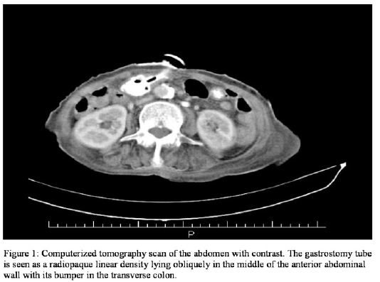
Computirized tomography confirmed misplacement, localizing the position of the bumper in the transverse colon (Figure-1). No contrast material entered the peritoneal cavity. Laparotomy was performed to close the fistula and to replace the gastrostomy (Figure-2). The postoperative course was uneventful, and she had no further difficulty with enteral feeding via the new gastrostomy tube.
Discussion
Percutaneous endoscopic gastrostomy tubes are a well-established, effective, and a relatively safe technique for long term feeding of patients. In general the complication rate is low and migration of a PEG tube into the colon originally positioned in the stomach is an extraordinarily rare complication, typically occurring within days to month after insertion (8, 9). It has also been found in patients with previous abdominal surgery (10). Feculent vomiting and profuse diarrhoea are the two most common symptoms of gastrocolocutaneous fistula, both being present in our patient. What was unusual in this case was the time of presentation of the gastro-colic fistula long time post-PEG insertion and has no previous surgery.
The exact mechanism of gastrocolocutaneous fistula is not well known. However, the most plausible theory is the interposition of the colon, usually the transvers colon, between the anterior abdominal wall and the gastric wall (11, 12). The guide needle passed from the skin into the colon and then entered the stomach. The PEG tube followed that needle retrograde through the stomach, first penetrated the colon, and then exited the skin. This hypothesis is supported by the abdominal computed tomography scan (Figure-1).
Nonoperative management strategies for gastrocolo cutaneous fistula have been published, including fibrin glue, nonsteroidal anti-inflammatory drug, parenteral nutrition, and intravenous cimetidine (13–17). However, this non-operative managements may not be success in all patients. Surgery is required for definitive management of gastrocolic fistula. We demonstared the succesful surgical procedure for this case.
Techniques using both transillumination and finger pressure as a guide to place the puncture site are useful for preventing this complication. Guidance by ultrasound or CT can be used selectively but may have limited benefit. Foutch et al, suggested that an aspirating syringe filled with saline could be used to identify the intervening colon between the skin and the stomach if air bubbles appeared in syringe prior to the endoscopic visualization of the needle in the gastric lumen (10).
Conclusion
In conclusion, gastrocolocutaneous fistula must be cosidered as a complication of PEG tube placement when patients develop a sudden onset of unexplained diarrhea after PEG tube feeding, and should confirm the diagnosis by a CT. Delayed onset or intermittent occurrence of symptoms does not rule out a gastrocolocutaneous fistula. To avoid this complication, patients in a high risk group have to undergo careful assessment before PEG.
Declarations
- Author contributions (CRediT)
- All authors contributed equally to all 14 CRediT roles (Conceptualization, Data curation, Formal analysis, Funding acquisition, Investigation, Methodology, Project administration, Resources, Software, Supervision, Validation, Visualization, Writing – original draft, Writing – review & editing).
- Funding
-
Funding information was not reported in the published version of this article.
- Conflicts of interest
- A conflict of interest statement was not reported in the published version of this article.
- Ethics approval
- Ethics approval details were not reported in the published version of this article.
- Data availability
- A data availability statement was not reported in the published version of this article.
References (17)
11 of 17 references are linked to their DOI.
- Gauderer MWL. Percutaneous endoscopic gastrostomy 20 years later: historical perspective. J Pediatr Surg 2001;36:217-9. DOI Crossref PubMed Google Scholar
- Sherwin P, Schrag S, Sharma S, et al. Complications related to percutaneous endoscopic gastrostomy (PEG) tubes. A comprehensive clinical review. J Gastrointestin Liver Dis 2007;16:407–18. PubMed Google Scholar
- Sasaki T, Fukumori D, Sakai K, Sato M, Ohmori H, Yamamoto F. The safety and feasibility of percutaneous endoscopic gastrostomy placement. Hepatogastroenterology 2004;51:1062–5. PubMed Google Scholar
- Friedmann R, Feldman H, Sonnenblick M. Misplacement of percutaneously inserted gastrostomy tube into the colon: report of 6 cases and review of the literature. JPEN J Parenter Enteral Nutr 2007;31:469 –76. DOI Crossref PubMed Google Scholar
- Pitsinis V, Roberts P. Gastrocolic fistula as a complication of percutaneous endoscopic gastrostomy. Eur J Clin Nutr 2003;57:876-8. DOI Crossref PubMed Google Scholar
- Sarsu SB, Karapur A. Ileal Perforation Caused by Typhoid Fever. Ulutas Med J. 2015; 1(3): 86-89. DOI Crossref PubMed Google Scholar
- Marcy PY, Magne N, Lacroix J, Bailet C. Late presentation of gastrocolic fistula after percutaneous fluoroscopic gastrostomy. JBR-BTR 2004;87:17-20. PubMed Google Scholar
- Huang SY, Levine MS, Raper SE. Gastrocolic fistula with migration of feeding tube into transverse colon as a complication of percutaneous endoscopic gastrostomy. AJR Am J Roentgenol 2005;184:S65-S66. DOI Crossref PubMed Google Scholar
- Lenzen H, Weismuller T, Bredt M, Bahr M. Education and imaging. Gastrointestinal: PEG feeding tube migration into colon;late manifestation. J Gastroenterol Hepatol 2012;27: 1254. DOI Crossref PubMed Google Scholar
- Foutch PG et al. Percutaneous endoscopic gastrostomy in patients with prior abdominal surgery: virtues of the safe tract. Am J Gastroenterol 1988;83:147-150. PubMed Google Scholar
- Roozrokh HC, Ripepi A, Stahlfeld K. Gastrocolocutaneous Fistula as a complication of peg tube placement. Surg Endosc. 2002;16:538–9. DOI Crossref PubMed Google Scholar
- Minocha A, Rupp TH, Jaggers TLSilent cologastrocutaneous fistula as complication of percutaneous endoscopic gastrostomy. Am J Gastroenterol 1994;89:2243-2244. PubMed Google Scholar
- Rots WI, Mokoena T. Successful endoscopic closure of a benign gastrocolonic fistula using human fibrin sealant through gastroscopic approach: a case report and review of the literature. Eur J GastroenterolHepatol 2003;15:1351-6. DOI Crossref PubMed Google Scholar
- Shand A, Reading S, Ewing J, et al. Palliation of a malignant gastrocolic fistula by endoscopic human fibrin sealant injection. Eur J GastroenterolHepatol 1997;9:1009-11. DOI Crossref PubMed Google Scholar
- Tan CC, Guan R et al. Closure of a benign gastrocolic fistula on medical management. Singapore Med J 1994;35:423-5. PubMed Google Scholar
- Thyssen EP, Weinstock LB, Balfe DM, et al. Medical treatment of benign gastrocolic fistula. Ann Intern Med 1993; 118:433-5. DOI Crossref PubMed Google Scholar
- Ahamed F, Chandrappa N, Abdallah EM. A comparative study of diagnosis methods for detection of Helicobacter pylori in gastro-duodenal diseases in Human. Ulutas Med J. 2015; 1(3): 64-67. Cite? DOI Crossref PubMed Google Scholar
Read the full text
Open the PDF here
Loading the PDF…
If the PDF does not show here, use the download button above.
How to cite this article
Sarici IS, Kala F, Kara G, Eskut N, Gundogdu E. Gastrocolocutaneous Fistula-Delayed complication of Percutaneous Endoscopic Gastrostomy, Case report. Ulutas Med J. 2015;1(4):116-118. doi:10.5455/umj.20150417122313
Sarici, I. S., Kala, F., Kara, G., Eskut, N., & Gundogdu, E. (2015). Gastrocolocutaneous Fistula-Delayed complication of Percutaneous Endoscopic Gastrostomy, Case report. The Ulutas Medical Journal, 1(4), 116–118. https://doi.org/10.5455/umj.20150417122313
Sarici, I.S. et al. (2015) 'Gastrocolocutaneous Fistula-Delayed complication of Percutaneous Endoscopic Gastrostomy, Case report', The Ulutas Medical Journal, 1(4), pp. 116–118. doi: 10.5455/umj.20150417122313.