The Ulutas Medical Journal · ISSN 2149-0430 (print), 2149-388X (online) · https://www.ulutasmedicaljournal.com/?mno=210730
Original Research · Open access
Right ventricle diastolic function in high-normal and mildly elevated arterial blood pressure
- Cardiovascular Department of Cairo University, Egypt ROR
Abstract
- Introduction
- Many studies have evaluated left ventricle (LV) function in mildly increased arterial blood pressure; however, the right ventricle (RV) has received little attention because of its complex shape and orientation. This study aimed to evaluate RV diastolic dysfunction using pulsed and tissue Doppler echocardiography in untreated patients with blood pressure (BP) values ranging from optimal to mild hypertension.
- Materials and Methods
- The study group consisted of 57 patients with high-normal and mildly increased arterial BP. Twenty healthy age- and sex matched subjects were recruited as controls. Conventional echocardiography was done to study LV dimensions and systolic function. Pulsed Doppler echocardiography was used to measure peak early and late tricuspid diastolic filling velocities. Tissue Doppler imaging was done to determine RV annular velocities.
- Results
- Right ventricular hypertrophy was found in 28% of our patients with newly diagnosed systemic hypertension (n=16). Tricuspid E/A was 1.3 and 0.9 in the control and hypertensive groups, respectively. Tissue Doppler tricuspid E/A was significantly decreased in the hypertensive group (0.8 ±0.2 versus 1.2 ±0.1 in control, p<0.001).
- Conclusions
- right ventricular diastolic function deteriorates with a slight increase in systemic blood pressure. Right ventricular diastolic dysfunction may be an early clue to hypertensive heart disease.
Keywords
Full text
Introduction
Arterial systemic hypertension may determine impairment of both left ventricle (LV) diastolic and systolic functions, due to increased after load (1). Also the right ventricle (RV) might be involved in this process by structural and functional abnormalities (2). Left ventricular hypertrophy is often associated with hypertension and is an adaptive mechanism to maintain or normalize wall stress, sometimes at the expense of diastolic and long-axis systolic function (3). This adaptive mechanism is associated with changes in left ventricular parameters and in chamber dimensions, geometry and function. These changes are progressive and can ultimately lead to heart failure with systolic and/or diastolic dysfunction (4). Human heart functions as a unit that includes the right ventricle. The spectrum of changes in structure, function and shape of the left ventricle ultimately has an effect on the structure and function of the RV (5).
Only a few publications concern echocardiographic assessment of the RV diastolic function in systemic hypertension, which is all more surprising as physical examination often demonstrates the features of RV pathology and dysfunction in hypertensive patients (6). Purpose of the study was assessment of RV diastolic function in patients with untreated systemic hypertension using conventional and doppler echocardiography.
Study Design
The study was carried out at the echocardiography laboratory of cardiovascular department at Cairo University hospitals. The Research Ethics Committee of the hospital reviewed and approved the study protocol. Fifty seven adult participants aged 54.8±14 years old with high-normal and mildly increased arterial blood pressure never treated with antihypertensive medications were compared with 20 age and gender matched control subjects .Patient written consent was given by all the participants. Hypertension was defined as persistent elevation of blood pressure above 140/90 mmHg on two or more occasions with the patient in a sitting position for at least five minutes (7). Patients were subjected to history taking, physical examination and simple clinical investigations. Demographic parameters, including age, gender and associated symptoms were obtained. Subjects with ischemic heart disease, valvular heart disease and cor pulmonale were all excluded.
Echocardiography
All subjects had echocardiography performed with the use of a Philips IE 33 (Andover, MA, USA) Ultrasound according to recommendations of the American Society of Echocardiography (8). Left ventricle structure, dimensions and functions were studied. Right ventricle wall thickness was measured in diastole from the sub costal views wall and wall thickness ≥ 5 mm indicates RV hypertrophy (9). Right ventricle diastolic function was assessed by recording tricuspid flow with standard pulsed wave Doppler and by measurements of early diastolic peak flow velocity (E), late diastolic peak flow velocity (A) and the ratio of early-to-late flow velocity peaks (E/A ratio).
Tissue Doppler imaging (TDI)
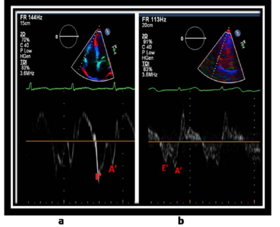
In the apical four-chamber view, the sample volume was placed at the lateral tricuspid annulus to study early diastolic (E') and late diastolic (A') velocities. The use of E':A' ratio is one of the recommended indices approved by the American Society of Echocardiography for assessing right ventricle diastolic dysfunction (8, 10).
Statistical analysis
All statistical calculations were done using computer program SPSS (Statistical Package for the Social Science, SPSS Inc., Chicago, IL, USA) release 15 for Microsoft Windows (2006). All Data were statistically described in terms of mean ± standard deviation (± SD) Comparison of numerical variables between the study groups was done using Student t test for independent samples and one way analysis of variance (ANOVA) test when comparing 3 group. P-value less than 0.05 was considered statistically significant.
Results
Fifty seven participants aged 54.8±14 years old (40 females and 17 males) with high-normal and mildly increased arterial blood pressure never treated with antihypertensive medications were compared with 20 age and gender matched control subjects free of any cardiac diseases. Systolic (SBP) and diastolic blood pressures (DBP) were significantly higher in hypertensive subjects compared to the controls (143.7±2.7 and 93.2±5.1 mmHg vs 119.4±8.1 and 72.3±6.9 mm hg, p-value <0.001 for both)(Table-1).

Conventional Echocardiography
Interventricular septum thickness and right ventricle free wall thickness were significantly increased in patients with mild untreated systemic hypertension (1.2±0.2 and 0.5±0.1 vs.0.8±0.1 cm and 0.28±0.1cm among control group, P<0.001for both).
Pulsed and tissue Doppler imaging
Mitral E/A ratio 1.2±0.1 and 0.7±0.1 in the control and hypertensive group respectively (P value < 0.001). While, tricuspid E/A was 1.3 and 0.9 in control and hypertensive groups respectively. Tissue Doppler tricuspid E' and E'/A' values were significantly decreased in hypertensive group (12±3.1 cm/s and 0.8 ±0.2 vs 15.3 ±1.3 cm/s and 1.2±0.1 in control group, p <0.001 for both) (Figure-1).Positive significant correlation was found between mitral and tricuspid E/A ratio (p<0.001, r: 0.842). Univariate regression analysis revealed that body mass index was found to be significant predictor of RV diastolic dysfunction (p:0.023).
Discussion
It was found that in middle-aged men central distribution of body fat is associated with increased BP, independently of body mass index and insulin resistance, thus suggesting a key role of central adiposity in the full expression of the “metabolic syndrome (11). The current study showed that body mass indices and waist circumferences were increased in the hypertensive group. Moreover, body mass index was found to be significant predictor of RV diastolic dysfunction (RVDD). This is concordant with other studies that found that mitral annular E':A' ratio and body mass index were the only predictors of RVDD (10). Concordant with other studies (12), the present study used the ratio E':A' ratio alone to assess RVDD . However, this index was approved by the American Society of Echocardiography to assess the global RV diastolic function. Positive significant correlation was found between mitral and tricuspid E/A ratio (p<0.001, r: 0.842) this is in accordance with other study that assessed RV DD in 100 adult Nigerian hypertensive subjects (13).
Concordant with some studies that found that the prevalence of RV hypertrophy consistently varied among studies (17.0-80.0%) with an average of 28.6% in the pooled population (14), the current study found that RV hypertrophy was prevalent in 28% of the high-normal and mildly increased blood pressure group. It was found that RV diastolic and systolic function deteriorates in response to slightly increased systemic BP (15). It was found that increased pulmonary vascular resistance in systemic hypertension results from the activation of vasoconstriction pathways in both systemic and pulmonary vascular beds which may explain right ventricular hypertrophy from mild to moderate, uncomplicated hypertensive patients (16).
Moreover, combination of increased thickness of the posterior wall, inter ventricular septum dimensions and right ventricular wall thickness will ultimately lead to at least progressive reduction in the right ventricular end-diastolic dimensions before progressive dilatation may occur in the right heart (17).
A magnetic resonance imaging study to assess RV remodeling in systemic hypertension revealed that that systemic hypertension was associated with concentric RV remodeling and impaired diastolic function, confirming that the unstressed ventricle is not immune to the effects of systemic hypertension (18).
Conclusion
In arterial hypertension, increased systemic after load is transferred to RV, a structure not directly exposed to systemic BP but the inter ventricular septum is likely the candidate for that role. Right ventricle diastolic function could provide novel insights in RV function impairment in high-normal and mildly increased arterial blood pressure. Moreover, RV diastolic dysfunction may be an early clue to hypertensive heart disease.
Declarations
- Author contributions (CRediT)
- All authors contributed equally to all 14 CRediT roles (Conceptualization, Data curation, Formal analysis, Funding acquisition, Investigation, Methodology, Project administration, Resources, Software, Supervision, Validation, Visualization, Writing – original draft, Writing – review & editing).
- Funding
-
No external source of funding was used for the current study.
- Conflicts of interest
- All authors declared no conflict of interest.
- Ethics approval
- The Research Ethics Committee of the hospital reviewed and approved the study protocol.
- Data availability
- A data availability statement was not reported in the published version of this article.
References (18)
11 of 18 references are linked to their DOI.
- Galderisi M, Petrocelli A, Alfieri A, Garofalo M, de Divitiis O. Impact of ambulatory blood pressure on left ventricular diastolic function. Am J Cardiology 1996, 77: 597– 601. PubMed Google Scholar
- Myslinski W, Mosiewicz J, Ryczak E, Barud W, Biłan A, Palusinski R´and Hanzlik J. Right ventricular function in systemic hypertension. J HumHypertension 1998, 12: 149-155. DOI Crossref PubMed Google Scholar
- Verdecchia P, Schillaci G, Borgioni C, et al. Prognostic value of left ventricular mass and geometry in systemic hypertension with left ventricular hypertrophy. Am J Cardiology 1996, 78: 197–202. DOI Crossref PubMed Google Scholar
- Janicki J. Influence of the pericardium and ventricular interdependence on left ventricular diastolic and systolic function in patients with heart failure. Circulation 1990, 81(suppl III): III- 15–III-20. PubMed Google Scholar
- Clyne C, Alpert J, Benotti J. Interdependence of the left and right ventricles in health and disease. Am Heart J 1989, 117: 1366–1373. DOI Crossref PubMed Google Scholar
- Cittadini A, Fazio S, Stromer H et al. Optimal determination of right ventricular filling dynamics in systemic hypertension. Am Heart J 1995, 130: 1074-1082. Noha Hassanin Hanboly et al. Right Ventricle Diastolic Function DOI Crossref PubMed Google Scholar
- Paul A, Suzanne O, Barry L, William C, Cheryl D, Joel H, Daniel T, et al. Report From the Panel Members Appointed to the Eighth Joint National Committee (JNC). JAMA 2014, 311:507-520. PubMed Google Scholar
- Lawrence G, Wyman W, Jonathan A, Lanqi H, Mark D, Krishnaswamy C, Scott D et al. Guidelines for the cardiography Assessment of the right heart in adults. J Am Soc cardiography 2010, 23:685-713. PubMed Google Scholar
- Roberto M, Luigi P, Victor M, Jonathan A, Anderson A, Laura E, Frank A et al. Recommendations for Cardiac Chamber Quantification by Echocardiography in Adults: An Update from the American Society of Echocardiography and the European Association of Cardiovascular imaging. J Am Soc Echocardiogr 2015; 28:1-39. DOI Crossref PubMed Google Scholar
- Cicala S, Galderisi M, Caso P, Petrocelli A, D’ Errico A, de Divitiis O et al. Right ventricular diastolic dysfunction in arterial systemic hypertension: analysis by pulsed tissue Doppler. Eur J Echocardiography 2002; 3:135– 142. DOI Crossref PubMed Google Scholar
- Alfonso S, Francesco P, Gianvincenzo B. The Relationship of waist circumference to blood pressure. AJH 2002,15:780-786. PubMed Google Scholar
- Kamilu M, Hadiza S, Mohammed N. Right ventricular dysfunction in a hypertensive population stratified by patterns of left ventricular geometry. Cardiovascular J Africa 2012; 23: 478–482. PubMed Google Scholar
- Akintunde A, Akinwusi P, Familoni O and Familoni O. Effect of systemic hypertension on right ventricular morphology and function: an echocardiography study.Cardiovasc J Afr. 2010 Oct; 21(5): 252– 256. DOI Crossref PubMed Google Scholar
- Cuspidi C, Sala C, Muiesan M, et al. Right ventricular hypertrophy in systemic hypertension: an updated review of clinical studies. J Hypertension 2013, 31:858-65. DOI Crossref PubMed Google Scholar
- Pedrinelli R, Canale M, Giannini C, Talini E, Penno G, Dell'Omo G, Di Bello V. Right ventricular dysfunction in early systemic hypertension: a tissue Doppler imaging study in patients with high-normal and mildly increased arterial blood pressure. J Hypertension 2010, 28:615 -21. DOI Crossref PubMed Google Scholar
- Vriz O, Argiento P, D'Alto M, Ferrara F, Vanderpool R, Naeije R, Bossone E. Increased pulmonary vascular resistance in early stage systemic hypertension: a resting and exercise stress echocardiography study. Can J Cardiol 2015; 4:537-43. DOI Crossref PubMed Google Scholar
- Nunez B, Amodeo C, Garavaglia G, Schmiedt R, Frohlich E. Biventricular cardiac hypertrophy in essential hypertensio n. Am Heart J 1987;114:813-818. PubMed Google Scholar
- Todiere G, et al. Right ventricular remodeling in systemic hypertension:a cardiac MRI study. Heart 2011, 97:1257-61. DOI Crossref PubMed Google Scholar
Read the full text
Open the PDF here
Loading the PDF…
If the PDF does not show here, use the download button above.
How to cite this article
Hanboly NH. Right ventricle diastolic function in high-normal and mildly elevated arterial blood pressure. Ulutas Med J. 2015;1(4):100-103. doi:10.5455/umj.20151225044952
Hanboly, N. H. (2015). Right ventricle diastolic function in high-normal and mildly elevated arterial blood pressure. The Ulutas Medical Journal, 1(4), 100–103. https://doi.org/10.5455/umj.20151225044952
Hanboly, N.H. (2015) 'Right ventricle diastolic function in high-normal and mildly elevated arterial blood pressure', The Ulutas Medical Journal, 1(4), pp. 100–103. doi: 10.5455/umj.20151225044952.