The Ulutas Medical Journal · ISSN 2149-0430 (print), 2149-388X (online) · https://www.ulutasmedicaljournal.com/?mno=134160
Original Article · Open access
Protective Effects of Micro-Injected Adipose-Derived Mesenchymal Stem Cells on Ovarian Ischemia/Reperfusion Injury in Rats
- Muğla Sıtkı Koçman Üniversitesi ROR
- Department of Histology-Embryology, School of Medicine, Celal Bayar University, Manisa, Turkey. ROR
- Department of Laboratory Animal Science, School of Medicine, Dokuz Eylul University, İzmir, Turkey. ROR
- Department of Clinical Biochemistry, School of Medicine, Mugla Sitki Kocman University, Mugla, 48000, Turkey ROR
- Department of Obstetrics and Gynecology, School of Medicine, Mugla Sitki Kocman University, Mugla, 48000, Turkey ROR
Abstract
- Introduction
- The authors investigated the immunohistochemical, histological, and biochemical effects of adipose-derived stem cells (ADSC) which possess antioxidant and anti-inflammatory properties in the ovarian ischemia/ reperfusion rat model.
- Materials and Methods
- Rats were randomly allocated to sham, ADSC, torsion/detorsion, torsion/detorsion/ADSC groups. ADSCs were micro-injected to torsion/detorsion/ADSC during the detorsion procedure after a 6-h period of torsion. BrdU, caspase-3, TUNEL, eNOS, and iNOS staining were used. Advanced oxidation protein products (AOPPs), superoxide dismutase (SOD) activity, and levels of total sulfhydryl (TSH) were evaluated.
- Results
- Serum AOPP increased in the torsion/detorsion, tissue and serum AOPP levels in the torsion/detorsion/ ADSC group were decreased. Tissue SOD activity in torsion/detorsion and torsion/detorsion/ADSC groups was significantly decreased compared to the sham group. The iNOS was stronger in the torsion/detorsion group following torsion and detorsion but decreased after ADSC microinjection in the torsion/detorsion/ADSC group. ADSC treatment after torsion-detorsion also reduced caspase-3 immunoreactivity and apoptotic cell number.
- Conclusion
- ADSC implementation after torsion/detorsion might protect the ovary from ischemia/reperfusion injury.
Keywords
Full text
Introduction
Torsion of the adnexa, the rotation of the fallopian tube and ovary, accounts for 2.7−3% of gynecological emergencies (1). Since most cases occur at reproductive ages, early diagnosis and effective treatment are vital for maintaining the ovarian reserve and hormonal functions. Salpingo-oophorectomy was the previously preferred management option due to risks of thromboembolism and potential malignancy with a nonviable appearance of the ovary; but recent studies recommend ovarian detorsion for patients desiring to protect their fertility (2). In ovarian torsion, gangrene and necrosis with venous and lymphatic occlusion and subsequent arterial insufficiency may ultimately cause loss of ovarian reserve due to ischemia (3). After reperfusion of the ovary by detorsion, many reactive oxygen species (ROS), namely hydroxyl radicals, hydrogen peroxide, and superoxide radicals develop due to increased oxygen levels (4, 5). The elevated ROS result in DNA damage, endothelial destruction, and apoptosis of granulosa cells (6, 7). To improve the outcomes of ovarian detorsion operations, many promising anti-inflammatory and antioxidant agents have been investigated such as atorvastatin, Galega officinalis, Olea europaea and enoxaparin(8–11). Human adipose-derived stem cells (ADSCs) are derived from human adipose tissue as a kind of superior biomaterial that can be suitable for allotransplantation and regenerative medicine (12). ADSCs stably expand under basic culture conditions, exhibit a low risk of contamination, and they have low immune rejection following transplantation (13). ADSCs also possess antioxidant and anti-inflammatory properties and have recently been used in many rat I/R models in the liver and brain (12, 14). Stem cells are also being investigated for obstetric and gynecological applications such as in Asherman syndrome, stress urinary incontinence, and premature ovarian failure (15–17). Furthermore, previous studies showed that cytokines secreted from ADSCs could resist the process of natural ovarian aging (18). Recently it has been demonstrated that human ADSC-derived exosomes recover the function of premature ovarian failure by increasing the proliferation rate and inhibiting the apoptosis rate of the human granulosa cells (19).
In this study, we aimed to investigate the effects of ADSCs in I/R injury caused by ovarian torsion/detorsion surgery.
Materials and Methods
Animals
A total of 25 adult, virgin, 10–12 weeks old female Wistar albino rats were allocated in this study. The animals (170−210 g) were housed under conditions of 20–24°C, 12-h of day/night cycles, and 50–60% humidity. The Dokuz Eylül University Ethics Committee for Animal Research approved the animal protocol for this study (Approval no. 28/2015). Animal research investigations conformed to the experimental guidelines in line with Dokuz Eylül University Committee for Human Care.
ADSC Isolation
ADSCs were isolated from five rats that were not allocated to experimental groups. Anesthesia was induced by intraperitoneal 10 mg/kg (Rompun, Bayer, Leverkusen, Germany) xylazine hydrochloride, and 50 mg/kg ketamine administration (Ketalar, Eczacibasi, Turkey). The antiseptic solution was used for surgical site preparation and the bilateral groin fat pads were removed. Adipose tissues were densely processed in sterile phosphate-buffered saline (PBS) with 5% penicillin-streptomycin.
Tissues were dissociated with scalpels and incubated for 30 min in α-MEM containing 0.1% collagenase Type 1 at 37°C in a 5% CO2. Tissues were then transferred to media for culture (20% fetal bovine serum (FBS), α-MEM, 1% L-glutamine), and after being centrifuged twice, they were cultivated in humidified incubators in α-MEM/10% FBS, 50 μg/ml streptomycin and 50 U/ml penicillin at 37°C and 5% CO2 (20). The media were changed every two days during the 10-day cultivation period.
Characterization of ADSCs
The cells were fixed in 4% paraformaldehyde for 30 minutes in PBS and permeabilized for 15 min with 0.1 Triton X-100 (A4975, AppliChem, Darmstadt, Germany after washing with PBS. Endogenous peroxidase activity of cells was put out by incubation with 3% hydrogen peroxide (K31355100, Merck, Darmstadt, Germany) for 5 min at room temperature. Afterward, cells were washed with PBS. After washing, cells were incubated in blocking serum for 1h in compliance with the manufacturer’s instructions (85-9043, Invitrogen, Camarillo, CA, USA). Anti-CD-34 (ab51037, Abcam) and anti-CD-44 (BA3382, BOSTER) primary antibodies were applied at a dilution of 1:25 and 4°C overnight (20). Primary antibodies were removed and cells were washed with PBS. Biotinylated goat IgG, anti-rabbit/mouse IgGsecondary antibody, and peroxidase-conjugated streptavidin (D01- 110, Life Science Division), were sequentially added, with each step incubated for 30 min. Cells were washed three times with PBS between these incubation periods. Then, the samples were incubated with diaminobenzidine /hydrogen peroxide (00-2020, Invitrogen) for 5 min. Mayer’s hematoxylin (HMM999, Scytek Laboratories, West Logan, Utah, USA) was used for counterstaining. The tissues were washed in distilled water, mounted with mounting medium (AML060, Scytek), and covered with coverslips for evaluation under the light microscope (Olympus BX40, Olympus Corp., Tokyo, Japan). The same procedures were applied for the control samples using the same type of IgGs instead of primary antibodies.
Treatment Groups
Animals were randomly distributed: group 1 (sham, saline n: 4), group 2 (ADSCs, n:7), group 3 (torsion + detorsion + saline, n:6), group 4 (torsion + detorsion + ADSCs, n:8).
Surgical Procedure
Rats underwent surgery in a supine position under general anesthesia as described in ADSC Isolation. After shaving and cleaning the surgical sites with antiseptic solution (Baticon, Drogsan, Turkey), 2.5-cm midline vertical incisions were made.
Group 1: Uterine corns and adnexa were visualized. Then, 20 µl saline was administered separately to the ovaries with a microinjector. Group 2: After exposing the adnexa, 1 x 10⁵ cells/ml ADSCs in 20 µl PBS (including 2000 cells of ADSCs) were separately administered to the ovaries with a microinjector (13, 21).
Group 3: To create torsion, the adnexa with connecting vessels were rotated 720° clockwise, and rotated adnexa were affixed to the anterior wall by 5/0 silk sutures. The abdominal wall incision was closed. Following a 6-h period of torsion, a re-laparotomy was performed for detorsion (reperfusion) by rotating the ovaries 720° counterclockwise and both ovaries were exposed. Then, 20 µl saline was administered separately.
Group 4: 6-h torsion and detorsion procedures were performed as described above. After the ovaries were exposed in the detorsion procedure, 1 x 10⁵ cells/ml ADSCs in 20 µl PBS (2000 cells of ADSCs) were administered separately with a microinjector (13, 21).
Abdominal wall incisions were all closed with 3/0 silk sutures. After surgical recovery, animals were housed separately. Following 7 days of the recovery period, all animals underwent laparotomy, and the oophorectomies were performed. All procedures were performed by the same researchers. One of the paired ovaries was selected randomly and fixed in 10% buffered formalin solution for histological and immunohistochemical analysis, while the other one was packed in oiled foil paper for further biochemical analysis. Just before sacrification of the rats via hypovolemic shock, 2 ml of blood was withdrawn from the inferior vena cava. After 15 min centrifugation of blood samples at 5000 rpm, the serum was collected in Eppendorf tubes. The Eppendorf tubes and packages of ovaries in oiled foil papers were transferred to dry ice and stored at -20°C until final biochemical analysis.
Histopathological Examination
Tissue samples were fixed for 48 hours in a 10% buffered formalin solution, dehydrated through alcohol, and inserted in paraffin. Paraffin blocks were sliced into sections with 5-µm-thickness (Leica RM2125RTS, Leica Biosystems, Nussloch, Germany). Hematoxylin and eosin staining was used as a routine protocol. The sections were examined for the severity of ovarian damage, such as edema, inflammation, hemorrhage, and congestion. All sections were examined under light microscopy (Olympus BX40, Tokyo, Japan) using the image analysis method (Image-Pro Plus 5.1). Tissue sections were analyzed by the same experienced histologist.
Immunohistochemical Analysis
For immunohistochemical analysis, Terminal deoxynucleotidyl transferase dUTP nick end labeling (TUNEL), Bromode oxyuridine/5- Bromo-2'-deoxyuridine (BrdU), caspase-3, Endothelial nitric oxide synthase (eNOS), and inducible nitric oxide synthase (iNOS) stainings were used. The intensity of immunolabeling was graded as follows: weak (+); moderate (++); and strong (+++). This grading was performed by two observers who were blinded to the experimental conditions.
TUNEL Assay
The slides were mounted with Entellan® (Merck 107961.0500) and examined by two histologists independently. 100 TUNEL- negative or positive- cells were counted per case on randomly chosen fields. The TUNEL-positive cells’ percentage was determined.
Biochemical Analysis
Ovarian tissues were homogenized in 20 mM phosphate buffer at pH 7.4. Homogenized samples were centrifuged at 14000 rpm at 40 C for 15 min. After centrifugation, supernatants were divided into aliquots for analysis of superoxide dismutase (SOD) activity, advanced oxidation protein products (AOPP), total sulfhydryl (TSH), and total protein levels, while pellets were used for the myeloperoxidase (MPO) activity.
Measurement of SOD Activity
The SOD activity of ovarian tissues or serum was measured spectrophotometrically (22). SOD activity in ovarian and serum were given as units/mg protein and units/mL, respectively.
Assay of TSH
The TSH levels of ovarian tissue or serum were evaluated with a linear standard curve formed by using reduced glutathione (23). Protein levels in supernatants with sodium dodecyl sulfate were measured according to Lowry et al (24). TSH levels were expressed as μmol/mg protein and μmol/L for tissue or serum respectively.
Assay of AOPP Levels
AOPP levels in ovarian tissue and serum were measured according to the spectrophotometric method of Witko-Sarsat (25).
Assay of MPO Activity
Bradley’s method was used for measuring MPO activity in the ovary (26).
Total Protein Assay
Total protein levels in ovarian tissue were measured according to the spectrophotometric method (27).
Statistical Analysis
The authors determined the sample size from the results obtained from the rats that they evaluated preliminarily. From the differences, a domineering two-tailed α value of 0.05 and a β value of 0.20 (study power: 80%). SPSS version 22.0 software (Chicago, IL, USA) was used. Distribution normality was assessed with the Shapiro–Wilk test. Results are given as mean ± standard error or median (min-maximum). Differences among experimental groups were determined by one-way analysis of variance (ANOVA) and Tukey’s post hoc tests. Kruskal– Wallis was used for analyzing non-parametric data. p<0.05 was considered significant.
Results
Results of ADSC Characterization
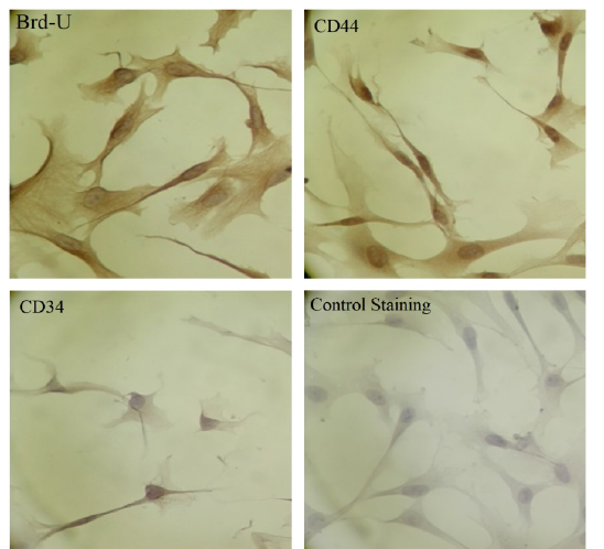
ADSCs were CD 34 (-) (negative) and CD 44 (+) (positive) (Fig.1). ADSCs were confirmed not to be hematopoietic stem cells as being CD34 (-) and confirmed to be adipogenic stem cells since they were CD44 (+).
Histological Results
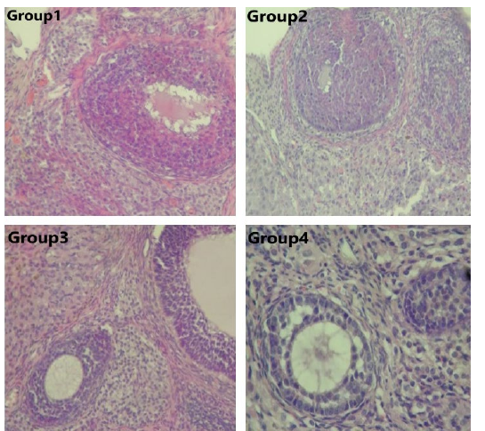
In the gross examination, the color of the ovaries in groups 1, 2, and 4 were less ischemic than group 3. Fibroblast accumulation around the ovarian follicles and small blood vessels filled with erythrocytes in the ovarian stroma were observed in group 1, whereas these changes decreased after ADSC administration in group 2. Intensely detected stromal fibroblast accumulation, infiltration and apoptotic cells in group 3 decreased after ADSC administration in group 4 than any other groups (Figure 2).
Immunohistochemical Results
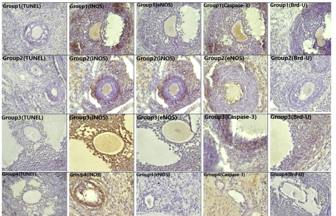
TUNEL analysis of ovarian tissue apoptosis is shown in Figure 3. No TUNEL-positive cells were observed in primary, secondary, or Graafian follicles in groups 1 and 2. However, apoptotic cells were detected in corona radiata and cumulus cells of Graafian follicles, and primary follicles of group 3. In group 4, TUNEL- positive cells were detected only in Graafian follicles, but in lower numbers than in group 3. Immunohistochemical distributions of iNOS, eNOS, Caspase-3, and BrdU are shown in Figure 3, respectively. TUNEL, iNOS, eNOS, Caspase-3, and BrdU staining levels in the groups are also demonstrated in Table 1. BrdU staining was positive only in groups 2 and 4, confirming the presence of ADSCs in ovarian tissue after stromal injection.

Biochemical Results
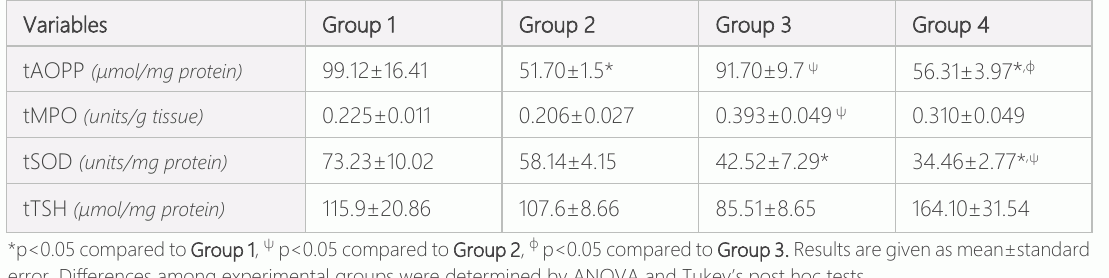
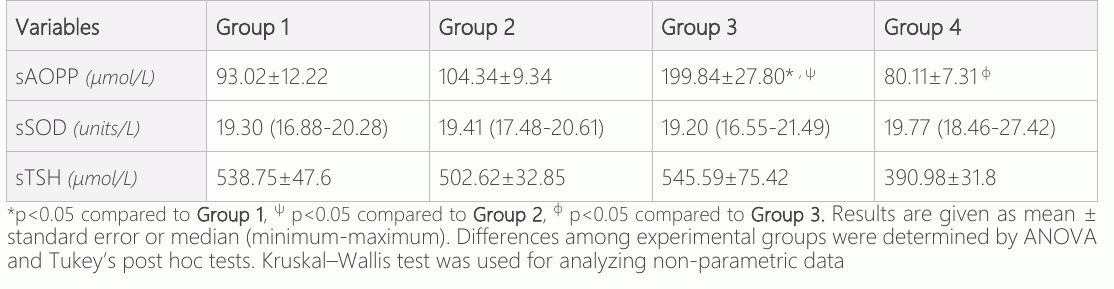
AOPP levels in tissue and serum of all groups are presented in Tables 2 and 3, respectively. Levels of tissue AOPP were higher in group 3 than in group 2 (p<0.05), whereas levels of AOPP in group 4 were lower than those in groups 1 and 3 (p<0.05) (Table 2). Serum AOPP was higher in group 3 than in group 1 and 2 (p<0.05), whereas they significantly decreased compared to group 3 after treatment with ADSCs in group 4 (p<0.05) (Table 3). SOD and MPO activities and TSH levels in tissue and serum in all groups are presented in Tables 2 and 3, respectively. Tissue SOD activity was significantly lower in group 3 than in group 1 (p<0.05). Additionally, we found that SOD activity in group 4 was significantly lower than that in groups 1 and 2 (p<0.05) (Table 2). The MPO activity in ovarian tissue in group 3 was greater than in group 2 (p<0.05 (Table 2).
Discussion
It was hypothesized that since ADSCs have antioxidant and anti-inflammatory properties, they might improve the harmful effects of I/R injury caused by ovarian torsion/detorsion surgery and the present study confirmed this by demonstrating that tissue and serum AOPP, as well as tissue SOD levels, were decreased after ADSC implementation. So, adjuvant treatment with ADSCs in an ovarian torsion model may mitigate ovarian damage by decreasing ROS in both tissue and serum, reducing the number of apoptotic cells and the inflammatory process. Many opinions have been reported regarding the effects and mechanism of ADSC on human infertility and the reproductive system recently. In a study by Manavella et al, ADSC differentiates into human vessels and promotes the secretion of VEGF when transplanted together with human ovarian tissue to mouse peritoneum using a two-step ovarian tissue grafting procedure (28). They stated that ADSC may potentially improve ovarian tissue quality and lifespan. In another of Manavella et al, authors demonstrated higher rates of oxygenation and vascularization of ovarian tissue with ADSC in the early post-grafting period, which correlate with increased follicle survival and reduced apoptosis (29). Moreover, Sun et al found that ADSC sheets transplanted into excised uteri promoted regeneration of the endometrium cells, muscle cells and stimulated angiogenesis, and also resulted in better pregnancy outcomes (30). As can be understood from these data, ADSCs appear to have positive effects on the human reproductive system and infertility.
The elevated levels of ROS could cause some damages to vital macromolecules including DNA, protein, and lipids and they play a significant role in the development of some pathological events such as I/R-induced ovarian injury (31). Such deleterious effects might be represented as a decrease in the number of follicles (pre-antral, and antral), as well as an increase in the number of atretic bodies and apoptosis of granulosa cells in follicles (10). The balance between ROS and antioxidants influences follicular growth and maturation because antioxidant supplements by reducing ROS and inhibiting lipid peroxidation can lead to preventing cell damage, enhancing follicular development, and increasing the fertilization rate and embryo development. The accumulation of ROS influences apoptosis. Shoorei et al found that hesperidin treatment led to an increase and a decrease in the mRNA levels of the Bcl-2 gene (anti-apoptotic) and Bax gene (pro-apoptotic), respectively. Hesperidin was also found to inhibit the generation of ROS. AOPPs have been widely used as reliable biomarkers for oxidant-mediated protein damage due to their stability (33). Previously, elevated plasma or serum AOPP has been reported to be associated with deterioration in atherosclerosis (34). In this study, AOPP was measured in both serum and tissue to estimate oxidant-mediated protein damage and conclusively it was found that ADSC treatment decreased the AOPP levels in ovarian tissue and serum following I/R injury. SOD and sulfhydryl groups are important antioxidants that protect the cell against free radical toxicity (35). Consistent with previous studies, decreased tissue SOD activity was found in the ovarian I/R model (35). Shojafar et al. performed bilateral oophorectomy/autotransplantation with and without ADSC implementation into autografted ovaries in mice to create an experimental model of I/R injury (36) They also presented the decrease of SOD in autografted ovaries and the increase of SOD in the group of autograft+ADSC. However, SOD activity was not significantly affected by treatment with ADSCs in current study. Besides, tissue TSH levels were decreased in the groups that underwent ovarian torsion compared to the control group 1, and TSH levels were higher in group 2 than in other groups. Nonetheless, these findings were not statistically significant. Additionally, there were no significant differences in systemic SOD and TSH levels among all the groups.
During I/R, increased ROS and other biological stimulants activate inflammatory cells, leading to neutrophil influx into the affected site. MPO, released by activated neutrophils, causes the formation of ROS and HClO (37). MPO activity was found to be significantly increased after ovarian I/R, but significant decrease in MPO activity after ADSC treatment was not observed. Histological analysis was consistent with this observation; lymphocyte infiltration gathered in group 3, and somewhat diminished following ADSC treatment.
Nitric oxide (NO) plays an essential role in vasodilation, immunity, inflammation and apoptosis. As a Janus molecule, NO employs both beneficial and deleterious effects depending on its isoforms, such as eNOS or iNOS (38). In this study, they found that ADSC treatment reduced iNOS expression in group 4 compared with 3. Several studies demonstrated that along with inflammation and apoptosis. Former researches revealed that the number of apoptotic cells was significantly increased in experimental models of ovarian I/R (8). In this study, apoptosis was evaluated immune histochemically with TUNEL and caspase-3 staining. Manavella et al. grafted frozen-thawed human ovarian tissue of five different patients in severe combined immunodeficient mice and intraperitoneally transplanted ADSC loaded in fibrin. They reported that significantly fewer apoptotic follicles were detected in the ADSC-loaded group in the TUNEL assay (29). In this study, a reduced number of apoptotic cells with ADSC treatment was shown by TUNEL assay. There are some similar studies published on testicular I/R injury. In the study of Moghimian et al. conducted on rats with testicular I/R injury, the level of testosterone increased and MDA level decreased significantly in the 5 h testicular torsion group receiving treatment with vitamin C. The authors concluded that this may be a result of the antioxidant effect of vitamin C (39). In the study of Shokoohi et al conducted on rats receiving hesperidin treatment, hesperidin increased the serum levels of GPx and SOD enzymes and decreased the heightened concentrations of MDA in testicular tissue. Moreover, the authors stated that hesperidin decreased Bax and increased Bcl-2 expression levels. They also demonstrated a markedly diminished rate of apoptosis in germ cells. The authors concluded that this may be a result of the antioxidant effect of hesperidin (40). In the study of Ameli et al conducted-on rats with testicular torsion, the levels of SOD, GPx, and testosterone were significantly decreased in the torsion/detorsion group while these increased in therapeutic groups receiving tadalafil and verapamil. Tadalafil and verapamil can protect the testis tissue damage and replace the testicular function by suppressing oxidative stress after testicular torsion (41). In the current study, the authors found that oxidative stress in ovarian tissue also increased after ischemia. Moreover, tissue SOD activity and apoptotic cell number were found to be significantly decreased in group 4. These findings might be the result of the antioxidant properties of ADSC, similar to the studies mentioned above.
In addition to investigations demonstrating the therapeutic efficacy of stem cells, tumor-tropic adipose stem cells have been shown to support tumor vasculature formation, and enhance ovarian cancer progression (42). The tumorigenic potential of ADSCs in breast cancer was investigated by Eterno et al and reported that ADSCs were not tumorigenic but they might possess a tumorigenic potential in presence of c-Met-expression (43). Thus, precautions are advised for the use of stem cells in benign gynecological disorders.
The power of our study might have been increased by using longer ischemia durations and by detecting ovarian reserve via follicular count, anti-Müllerian hormone, inhibin B, estradiol, or fecundity. However, the presence of only the ADSC-given group is a strong side of our study in terms of seeing ADSC effects on the undamaged ovary.
In conclusion, if administered within 6 hours of torsion, ADSC treatment might protect against ovarian I/R damage by decreasing protein oxidation and iNOS expression, attenuating lymphocyte infiltration, and decreasing the incidence of apoptotic cells. According to the results of the present study, a promising aspect of ADSCs might have lightened for human studies and finally, ADSCs can be used as a potential adjuvant treatment agent in ovarian torsion cases.
Declarations
- Author contributions (CRediT)
- All authors contributed equally to all 14 CRediT roles (Conceptualization, Data curation, Formal analysis, Funding acquisition, Investigation, Methodology, Project administration, Resources, Software, Supervision, Validation, Visualization, Writing – original draft, Writing – review & editing).
- Funding
-
Funding information was not reported in the published version of this article.
- Conflicts of interest
- The authors declared no conflict of interest.
- Ethics approval
- The Dokuz Eylül University Ethics Committee for Animal Research approved the animal protocol for this study (Approval no. 28/2015).
- Data availability
- A data availability statement was not reported in the published version of this article.
References (43)
38 of 43 references are linked to their DOI.
- Kart C, Aran T, Guven S, Karahan SC, Yulug E. Acute increase in plasma D- dimer level in ovarian torsion: An experimental study. Human Reproduction. 2011;26(3): 564–568 DOI Crossref PubMed Google Scholar
- Taskin O, Birincioglu M, Aydin A, Buhur AU, Burak F, Yilmaz I, Wheeler JM. The effects of twisted ischaemic adnexa managed by detorsion on ovarian viability and histology: An ischaemia-reperfusion rodent model. Human Reproduction. 1998; 13(10): 2823–2827 DOI Crossref PubMed Google Scholar
- Soltani M, Moghimian M, Abtahi H, S. M. The protective effect of Matricaria chamomilla extract on histological damage and oxidative stress induced by Torsion/Detorsion in adult rat ovary. International Journal of Womens Health and Reproduction Sciences. 2017;5(3): 187–192 DOI Crossref PubMed Google Scholar
- Sezgin B, Kinci MF, Pirinççi F, Camuzcuoğlu A, Erel Ö, Neşelioğlu S, Camuzcuoğlu H. Thiol-disulfide status of patients with cervical cancer. Journal of Obstetrics and Gynaecology Research. 2020;46(11):2423-2429 PubMed Google Scholar
- Sezgin B, Pirinççi F, Camuzcuoğlu A, Erel Ö, Neşelioğlu S,Camuzcuoğlu H. Assessment of thiol disulfide balance in early‐stage endometrial cancer. Journal of Obstetrics and Gynaecology Research. 2020;46(7):1140-1147 DOI Crossref PubMed Google Scholar
- Gharamaleki H, Parivar K, Soleimani Rad J, Roshangar L, Shariati M. Effects of electromagnetic field exposure during the prenatal period on biomarkers of oxidative stress and pathology of testis and testosterone level of adult rats in F1 generation. Acta Endocrinologica. 2014; 10(4): 577–587 DOI Crossref PubMed Google Scholar
- Sapmaz Metin M, Topcu Tarladacalisir Y, Uz YH, Inan M, Omurlu IK, Cerkezkayabekir A, Kizilay G, Akpolat M. Vitamin E modulates apoptosis and c-jun N- terminal kinase activation in ovarian torsion-detorsion injury. Experimental and Molecular Pathology. 2013; 95(2): 213–219 DOI Crossref PubMed Google Scholar
- KayaC., Turgut H, Cengiz H, Turan A, Ekin M, Yaşar L. Effect of detorsion alone and in combination with enoxaparin therapy on ovarian reserve and serum antimüllerian hormone levels in a rat ovarian torsion model. Fertility and Sterility. 2014;102(3):878-884 246 77 Stem Cells and Ovarian Torsion Kasap et al. DOI Crossref PubMed Google Scholar
- Parlakgumus HA, Aka Bolat F, Bulgan Kilicdag E, Simsek E, Parlakgumus A. Atorvastatin for ovarian torsion: Effects on follicle counts, AMH, and VEGF expression. European Journal of Obstetrics and Gynecology and Reproductive Biology. 2014;175: 186–190 DOI Crossref PubMed Google Scholar
- Shokoohi M, Soltani M, Abtahi-Eivary SH, Niazi V, Poor M, Ravaei H, Salimnejad R, Moghimian M, Shoorei H. Effect of hydro-alcoholic extract of Olea europaea on apoptosis-related genes and oxidative stress in a rat model of torsion/detorsion-induced ovarian damage. Asian Pacific Journal of Reproduction. 2019;8(4): 148–156 DOI Crossref PubMed Google Scholar
- Shokri F, Shokoohi M, Abadi ARR, Kalarestaghi H. The ameliorative effect of galega officinalis extract on histological damages, oxidative stress induced by torsion-detorsion in adult rats’ ovarian. International Journal of Women’s Health and Reproduction Sciences. 2019; 7(1): 119–123 DOI Crossref PubMed Google Scholar
- Yuk JS, Kim LY, Shin JY, Choi DY, Kim TY, Lee JH. A national population-based study of the incidence of adnexal torsion in the Republic of Korea. International Journal of Gynecology and Obstetrics. 2015;129(2): 169–170 DOI Crossref PubMed Google Scholar
- Takehara Y, Yabuuchi A, Ezoe K, Kuroda T, Yamadera R, Sano C, Murata N, Aida T, Nakama K, Aono F, Aoyama N, Kato K, Kato O. The restorative effects of adipose-derived mesenchymal stem cells on damaged ovarian function. Laboratory Investigation. 2013;93(2): 181–193 DOI Crossref PubMed Google Scholar
- Liu XL, Zhang W, Tang SJ. Intracranial transplantation of human adipose-derived stem cells promotes the expression of neurotrophic factors and nerve repair in rats of cerebral ischemia-reperfusion injury. International Journal of Clinical and Experimental Pathology. 2013;7(1): 174 –183 PubMed Google Scholar
- Gabr H, Rateb MA, El Sissy MH, Ahmed Seddiek H, Ali Abdelhameed Gouda S. The effect of bone marrow-derived mesenchymal stem cells on chemotherapy induced ovarian failure in albino rats. Microscopy Research and Technique. 2016;79(10): 938–947 DOI Crossref PubMed Google Scholar
- Kilic S, Yuksel B, Pinarli F, Albayrak A, Boztok B, Delibasi T. Effect of stem cell application on Asherman syndrome, an experimental rat model. Journal of Assisted Reproduction and Genetics. 2014.;31(8): 975– 982 DOI Crossref PubMed Google Scholar
- Wang Y, Shi G-wei, Wang H, Cao N-long, Fu Q. Adipose-derived stem cells seeded on polyglycolic acid for the treatment of stress urinary incontinence. World Journal of Urology. 2016;34(10): 1447–1455 DOI Crossref PubMed Google Scholar
- Su J, Ding L, Cheng J, Yang J, Li X, Yan G, Sun H, Dai J, Hu Y. Transplantation of adipose -derived stem cells combined with collagen scaffolds restores ovarian function in a rat model of premature ovarian insufficiency. Human Reproduction. 2016;31(5): 1075–1086 DOI Crossref PubMed Google Scholar
- Huang B, Lu J, Ding C, Zou Q, Wang W, Li H. Exosomes derived from human adipose mesenchymal stem cells improve ovary function of premature ovarian insufficiency by targeting SMAD. Stem Cell Research and Therapy. 2018;9(1): DOI Crossref PubMed Google Scholar
- Kurt O, Vatansever HS. Potential Clinical Use of Differentiated Cells From Embryonic or Mesencyhmal Stem Cells in Orthopaedic Problems. Current Stem Cell Research & Therapy. 2016;11(6): 522–529 PubMed Google Scholar
- Zhu SF, Hu HB, Xu HY, Fu XF, Peng DX, Su WY, He YL. Human umbilical cord mesenchymal stem cell transplantation restores damaged ovaries. Journal of Cellular and Molecular Medicine. 2015;19(9): 2108–2117 DOI Crossref PubMed Google Scholar
- Sun Y, Oberley LW, Li. A simple method for clinical assay of superoxide dismutase. Clinical Chemistry. 1988;34(3):497-500 DOI Crossref PubMed Google Scholar
- Taylan E, Resmi H. The Analytical Performance of a Microplate Method for Total Sulfhydryl Measurement in Biological Samples. Turkish Journal of Biochemistry. 2010;35(3): 275– 278 PubMed Google Scholar
- Lowry Oh, Rosebrough Nj, Farr AlRR. Protein measurement with the Folin phenol reagent. J Biol Chem. 1951;193(1):265- DOI Crossref PubMed Google Scholar
- Witko-Sarsat V, Friedlander M, Capeillère-Blandin C, Nguyen- Khoa T, Nguyen AT, Zingraff J, Jungers P, Descamps- Latscha B. Advanced oxidation protein products as a novel marker of oxidative stress in uremia. Kidney International. 1996;49(5): 1304–1313 DOI Crossref PubMed Google Scholar
- Bradley PP, Priebat DA, Christensen RD, Rothstein G. Measurement of cutaneous inflammation: Estimation of neutrophil content with an enzyme marker. Journal of Investigative Dermatology. 1982;78(3): 206–209 DOI Crossref PubMed Google Scholar
- Koike K, Moore FA, Moore EE, Trew CE, Banerjee A, Peterson VM. Endotoxin pretreatment inhibits neutrophil proliferation and function. Journal of Surgical Research. 1994;57(1): 49–54 DOI Crossref PubMed Google Scholar
- Manavella DD, Cacciottola L, Payen VL, Amorim CA, Donnez J, Dolmans MM. Adipose tissue-derived stem cells boost vascularization in grafted ovarian tissue by growth factor secretion and differentiation into endothelial cell lineages. Molecular Human Reproduction. 2019;25(4):184–193 DOI Crossref PubMed Google Scholar
- Manavella DD, Cacciottola L, Pommé S, Desmet CM, Jordan BF, Donnez J, Amorim CA, Dolmans MM. Two -step transplantation with adipose tissue-derived stem cells increases follicle survival by enhancing vascularization in xenografted frozen-thawed human ovarian tissue. Human Reproduction. 2018;33(6): 1107–1116 DOI Crossref PubMed Google Scholar
- Sun H, Lu J, Li B, Chen S, Xiao X, Wang J, Wang J, Wang X. Partial regeneration of uterine horns in rats through adipose - derived stem cell sheets. Biology of Reproduction. 2018; 99(5): 1057–1069 DOI Crossref PubMed Google Scholar
- Li C, Jackson RM. Reactive species mechanisms of cellular hypoxia- reoxygenation injury. In American Journal of Physiology - Cell Physiology. 2002; 282(2): 51-52 DOI Crossref PubMed Google Scholar
- Shoorei H, Banimohammad M, Kebria MM, Afshar M, Taheri MMH, Shokoohi M, Farashah MSG, Eftekharzadeh M, Akhiani O, Gaspar R, Pazoki-Toroudi H. Hesperidin improves the follicular development in 3D culture of isolated preantral ovarian follicles of mice. Experimental Biology and Medicine. 2019;244(5): 352–361 DOI Crossref PubMed Google Scholar
- Massy ZA, Nguyen-Khoa T. Oxidative stress and chronic renal failure: Markers and management. Int Journal of Nephrology. 2002;15(4): 336– 341 PubMed Google Scholar
- Liang X, Chen Y, Zhuang J, Zhang M, Xiong W, Guo H, Jiang F, Hu P, Guo D,Shi W. Advanced oxidation protein products as prognostic biomarkers for recovery from acute kidney injury after coronary artery bypass grafting. Biomarkers. 2012;17(6):507–512 DOI Crossref PubMed Google Scholar
- Zelko IN, Mariani TJ, Folz RJ. Superoxide dismutase multigene family: A comparison of the CuZn-SOD (SOD1), Mn-SOD (SOD2), and EC-SOD (SOD3) gene structures, evolution, and expression. In Free Radical Biology and Medicine. 2002;33(3): 337–349 247 77 Stem Cells and Ovarian Torsion Kasap et al. DOI Crossref PubMed Google Scholar
- Shojafar E, Soleimani Mehranjani M, Shariatzadeh SMA. Adipose derived mesenchymal stem cells improve the structure and function of autografted mice ovaries through reducing oxidative stress and inflammation: A stereological and biochemical analysis. Tissue and Cell. 2019.;56: 23–30. DOI Crossref PubMed Google Scholar
- Hampton MB, Kettle AJ, Winterbourn CC. Inside the neutrophil phagosome: Oxidants, myeloperoxidase, and bacterial killing. In Blood. 1998;92(9): 3007–3017 DOI Crossref PubMed Google Scholar
- Yang SR, Rahman I, Trosko JE., Kang KS. Oxidative stress-induced biomarkers for stem cell-based chemical screening. In Preventive Medicine. 2012;54:s 42–49 DOI Crossref PubMed Google Scholar
- Moghimian M, Soltani M, Abtahi H, Shokoohi M. Effect of vitamin C on tissue damage and oxidative stress following tunica vaginalis flap coverage after testicular torsion. Journal of Pediatric Surgery. 2017;52(10): 1651–1655 DOI Crossref PubMed Google Scholar
- Shokoohi M, Khaki A, Shoorei H, Khaki AA, Moghimian M, Abtahi-Eivary SH. Hesperidin attenuated apoptotic-related genes in testicle of a male rat model of varicocoele. Andrology. 2020; 8(1): 249–258 DOI Crossref PubMed Google Scholar
- Ameli M, Hashemi MS, Moghimian M, Shokoohi M. Protective effect of tadalafil and verapamil on testicular function and oxidative stress after torsion/detorsion in adult male rat. Andrologia. 2018;50(8): e13068 DOI Crossref PubMed Google Scholar
- Chu Y. Tang, Guo Y, Guo J, Huang B, Fang F, Cai J, Wang Z. Adipose-derived mesenchymal stem cells promote cell proliferation and invasion of epithelial ovarian cancer. Experimental Cell Research. 2015; 337(1): 16–27 DOI Crossref PubMed Google Scholar
- Eterno V, Zambelli A, Pavesi L, Villani L, Zanini V, Petrolo G, Manera S, Tuscano A, Amato A. Adipose -derived mesenchymal stem cells (ASCs) may favour breast cancer recurrence via HGF/c-Met signaling. Oncotarget. 2014;5(3): 613–633 DOI Crossref PubMed Google Scholar
Respond to this article
Comments on a published article are welcome as a Letter to the Editor.
Get an e-mail when new articles in your fields are published
Read the full text
Open the PDF here
Loading the PDF…
If the PDF does not show here, use the download button above.
How to cite this article
Kasap B, Kasap S, Vatansever S, Kendirci R, Yilmaz O, Calisir M, et al. Protective Effects of Micro-Injected Adipose-Derived Mesenchymal Stem Cells on Ovarian Ischemia/Reperfusion Injury in Rats. Ulutas Med J. 2021;7(4):237-247. doi:10.5455/umj.20211019101154
Kasap, B., Kasap, S., Vatansever, S., Kendirci, R., Yilmaz, O., Calisir, M., Turkcu, U. O., Akin, M. N., Akbaba, E., & Turhan, N. O. (2021). Protective Effects of Micro-Injected Adipose-Derived Mesenchymal Stem Cells on Ovarian Ischemia/Reperfusion Injury in Rats. The Ulutas Medical Journal, 7(4), 237–247. https://doi.org/10.5455/umj.20211019101154
Kasap, B. et al. (2021) 'Protective Effects of Micro-Injected Adipose-Derived Mesenchymal Stem Cells on Ovarian Ischemia/Reperfusion Injury in Rats', The Ulutas Medical Journal, 7(4), pp. 237–247. doi: 10.5455/umj.20211019101154.