The Ulutas Medical Journal · ISSN 2149-0430 (print), 2149-388X (online) · https://www.ulutasmedicaljournal.com/?mno=8877
Letter to the Editor · Open access
Pelvic Actinomycosis Associated with Intrauterine Device
- Division of General Surgery, Department of Surgery, Chi Mei Medical Center, Liouying, Tainan, Taiwan ROR
- Department of Obstetrics and Gynecology, Chi Mei Medical Center, Liouying, Tainan, Taiwan ROR
- Department of Pathology, Chi Mei Medical Center, Liouying, Tainan, Taiwan ROR
- Department of Intensive Care Medicine, Chi Mei Medical Center, Liouying, Tainan, Taiwan ROR
Abstract
Actinomycosis caused by Actinomyces genus is a subacute or chronic suppurative disease.1 Pelvic actinomycosis is usually associated with the longstanding use of IUDs.2 Because the clinical manifestation of pelvic actinomycosis is non-specific, it is hard to be differentiated actinomycosis from tuberculosis, malignancy, or another pelvic inflammatory disease. Thus, the preoperative diagnosis of pelvic actinomycosis is quietly difficult to reach. However, the clinician should keep alert this clinical entity in the presence of pseudotumoral mesentery infiltration on CT scan, especially for women with a longstanding IUD. The outcome of pelvic actinomycosis is favorable if the patient receiving the combinations of surgery and antibiotic treatment.
Keywords
Full text
Dear Editor, A 65-year-old woman presented with an abdominal pain for one month. In addition, she had poor appetite and body weight loss later. She had type 2 DM with poor control and had an intrauterine device (IUD) for 20 years. Physical examination was unremarkable except lower abdominal tenderness.
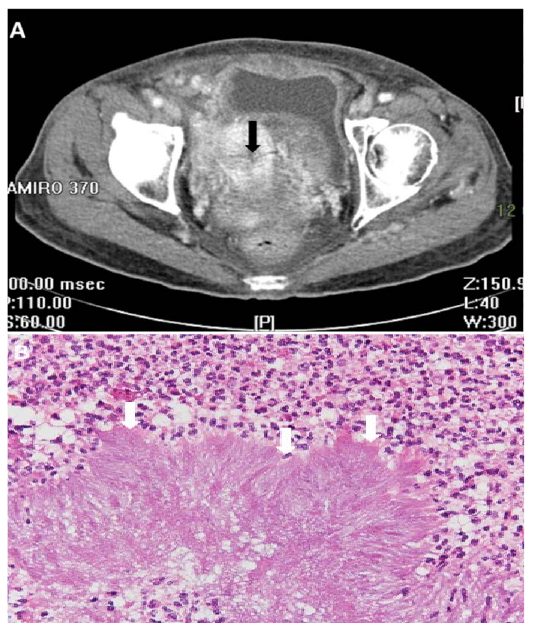
Colonoscopy showed partial obstruction at sigmoid colon due to suspicious external compression. Computed tomography image of abdomen revealed inflammatory change of pelvis with multi-focal abscess around a non-radiopaque IUD (Figure 1A, arrow). Exploratory laparotomy was performed, and severe ileum adhesion around IUD in pelvis was found. Loop ileostomy was undertaken after removal of IUD. The pathologic examination of implant and peritoneal nodules revealed an organized aggregate of filamentous bacteria surrounded by suppurative inflammation and a few sulfur granules, which indicated actinomyces infection (Figure 1B). Therefore, she received amoxicillin/clavulanate for five months, and the clinical condition recovered well. Finally, she received intestinal transit reconstruction after 6 months. Actinomycosis caused by Actinomyces genus is a subacute, or chronic suppurative disease (1). Pelvic actinomycosis is usually associated with the longstanding use of IUDs (2). Because the clinical manifestation of pelvic actinomycosis is non-specific, it is hard to be differentiated actinomycosis from tuberculosis, malignancy, or other pelvic inflammatory disease. Thus, preoperative diagnosis of pelvic actinomycosis is quietly difficult to reach. However, clinician should keep alert clinical entity in the presence of pseudotumoral mesentery infiltration on CT scan, especially for women with a longstanding IUD. The outcome of pelvic actinomycosis is favorable if patient receiving the combinations of surgery and antibiotic treatment (2).
Declarations
- Author contributions (CRediT)
- All authors contributed equally to all 14 CRediT roles (Conceptualization, Data curation, Formal analysis, Funding acquisition, Investigation, Methodology, Project administration, Resources, Software, Supervision, Validation, Visualization, Writing – original draft, Writing – review & editing).
- Funding
-
Funding information was not reported in the published version of this article.
- Conflicts of interest
- The authors declare that they have no conflict of interest in the letter .
- Ethics approval
- Ethics approval details were not reported in the published version of this article.
- Data availability
- Data sharing is not applicable to this article as no new data were created or analysed in this study.
References (2)
- Valour F, Sénéchal A, Dupieux C, et al. Actinomycosis: etiology, clinical features, diagnosis, treatment, and management. Infect Drug Resist 2014;7:183-197. PubMed Google Scholar
- García- García A, Ramírez-Durán N, Sandoval-Trujillo H, et al. Pelvic Actinomycosis. Can J Infect Dis Med Microbiol 2017;2017:9428650. PubMed Google Scholar
Respond to this article
Comments on a published article are welcome as a Letter to the Editor.
Get an e-mail when new articles in your fields are published
Read the full text
Open the PDF here
Loading the PDF…
If the PDF does not show here, use the download button above.
How to cite this article
Chuang CH, Huang SC, Lu NM, Chao CM. Pelvic Actinomycosis Associated with Intrauterine Device. Ulutas Med J. 2019;5(1):93-94. doi:10.5455/umj.20190919013918
Chuang, C. H., Huang, S. C., Lu, N. M., & Chao, C. M. (2019). Pelvic Actinomycosis Associated with Intrauterine Device. The Ulutas Medical Journal, 5(1), 93–94. https://doi.org/10.5455/umj.20190919013918
Chuang, C.H. et al. (2019) 'Pelvic Actinomycosis Associated with Intrauterine Device', The Ulutas Medical Journal, 5(1), pp. 93–94. doi: 10.5455/umj.20190919013918.